
Periprosthetic Joint Infection (PJI) Clinical Signs and Urgent Triage in Knee Arthroplasty
In short
Recognize the critical early warning signs of periprosthetic joint infection after knee replacement and master the urgent clinical triage protocol to protect the prosthesis.
In this guide (4 sections)
Periprosthetic joint infection (PJI) represents the single most dreaded and devastating complication following total knee arthroplasty, occurring in approximately one to two percent of primary procedures. Unlike standard soft tissue infections, bacteria that colonize prosthetic metallic and polyethylene surfaces rapidly secrete an extracellular polymeric substance, forming an impenetrable protective biofilm. Once a mature biofilm is established by organisms such as Staphylococcus aureus or coagulase-negative staphylococci, systemic antibiotics cannot eradicate the bacteria, frequently necessitating two-stage revision surgery with joint debridement, temporary antibiotic spacer placement, and prolonged disability. Early detection during the acute postoperative window (within the first four weeks) offers the only realistic opportunity for successful component retention through urgent surgical debridement and implant retention (DAIR). Every clinician and home caregiver must maintain aggressive vigilance for early signs of infection.
Environmental Dust, Tropical Perspiration, and Home Dressing Vulnerabilities
In India, managing post-operative wound hygiene involves specific environmental risk factors. Warm ambient temperatures, seasonal monsoons, and high humidity promote heavy bodily perspiration, which can seep beneath surgical dressings and macerate wound edges. Furthermore, environmental dust and the presence of family pets in living quarters present persistent contamination vectors. A frequent and perilous domestic error occurs when well-meaning relatives change surgical dressings using unsterilized bare hands, or wash the incision with tap water. Domestic caregivers must be trained in strict sterile wound protocols: dressings must remain dry, waterproof barriers should be used during sponge baths, and any damp or soiled bandage must be changed under aseptic conditions without touching the wound bed.
Biofilm Pathogenesis, Inflammatory Biomarkers, and Diagnostic Aspiration
The pathophysiology of acute PJI revolves around bacterial adherence to foreign implant biomaterials. Within hours of inoculation, planktonic bacteria adhere to titanium, cobalt-chromium, or polyethylene surfaces using surface protein adhesins. Once attached, they undergo phenotypic switching, encasing themselves in a dense biofilm matrix that blunts host neutrophil phagocytosis and elevates the minimum inhibitory concentration (MIC) of antibiotics by up to 1,000-fold. Differentiating normal postoperative inflammation from acute sepsis is critical. While mild incisional warmth and serous discharge are normal in the initial 72 hours, persistent wound drainage beyond day five, spreading erythema extending more than two centimetres from the incision line, new-onset joint effusion with severe resting pain, and systemic pyrexia indicate PJI. Immediate joint aspiration for synovial fluid leukocyte count, polymorphonuclear (PMN) percentage, and microbial culture is mandatory before any empirical antibiotics are initiated.
Immediate Cessation of Exercises, Non-Weight-Bearing Status, and Surgical Triage
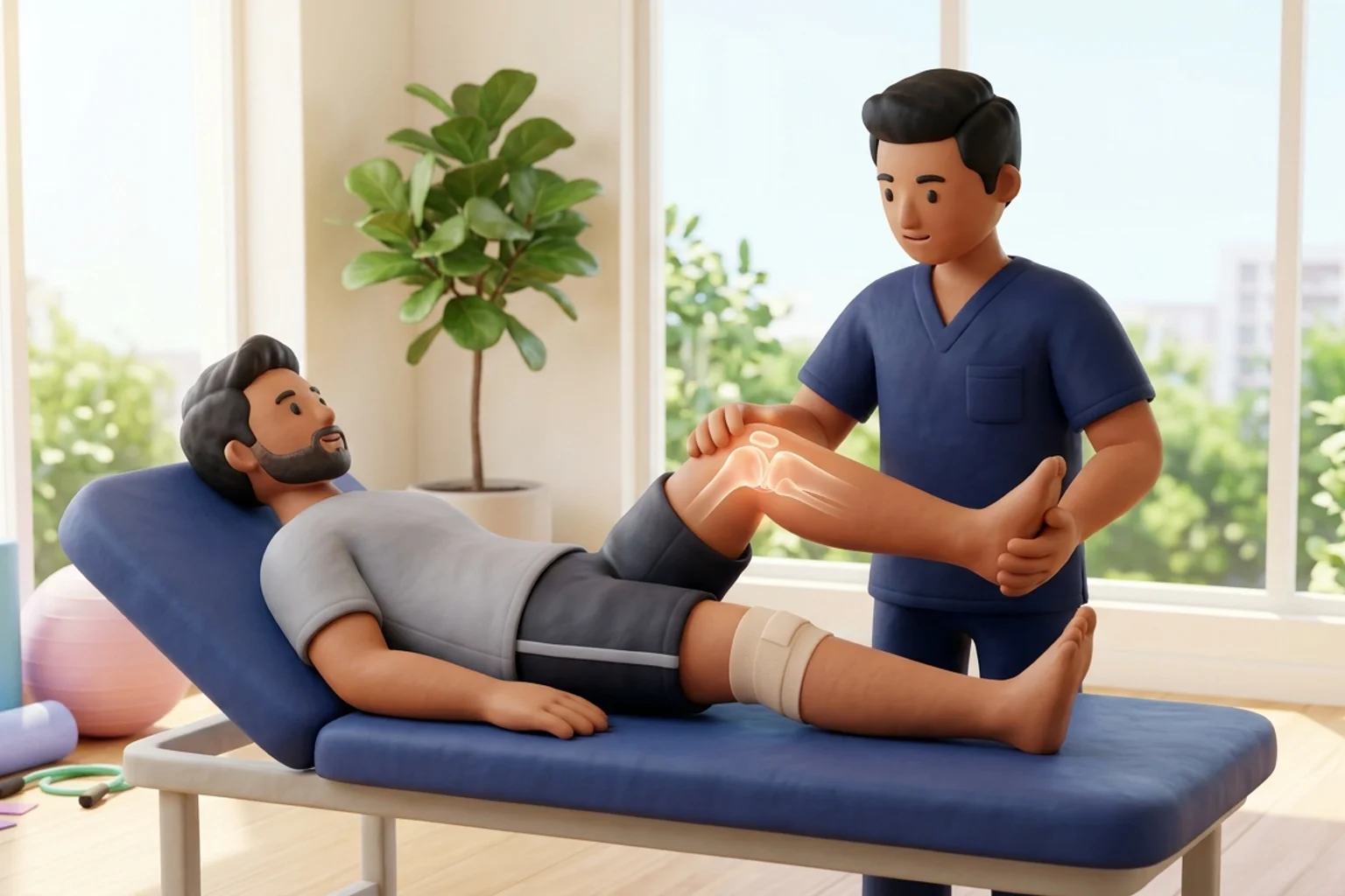
When clinical signs of PJI emerge, the role of the physiotherapist instantly transforms from active rehabilitation to urgent medical triage. All vigorous flexion-extension exercises, passive stretching, and resistance training must be halted immediately to avoid dispersing bacterial colonies or exacerbating joint capsular distension. The patient is placed on protected, non-weight-bearing mobility using a walker, the operative limb is immobilized in a comfortable extension splint, and urgent same-day referral to the operating orthopedic surgeon is executed. Physiotherapists must strictly resist the dangerous temptation to advise over-the-counter oral antibiotics, which suppress synovial fluid cultures without sterilizing the biofilm and obscure definitive diagnosis.
Protected Non-Weight-Bearing Bed Exercises During Clinical Evaluation
Objective: Maintain minimal calf circulation and prevent venous stasis while strictly avoiding mechanical stress on an inflamed joint
- 1Remain resting comfortably in a semi-reclined or supine position in bed with the surgical knee supported in gentle extension.
- 2Gently move both feet forward and backward at the ankles (light ankle pumps) through a comfortable, pain-free range.
- 3Perform gentle static gluteal contractions by squeezing the buttock muscles together for 3 seconds, then relaxing.
- 4Do not bend the knee, do not perform heel slides, and do not attempt to lift the straight leg against gravity.
- 5Ensure the limb remains elevated on a soft pillow while awaiting immediate surgical assessment.
| Clinical Parameter | Normal Post-Operative Healing Phase | Suspected Acute Periprosthetic Joint Infection (PJI) |
|---|---|---|
| Incisional Drainage | Minimal serosanguinous oozing resolving completely by post-op Day 3 to 4 | Persistent, expanding serosanguinous, cloudy, or frankly purulent drainage beyond Day 5 |
| Periwound Skin Changes | Mild, symmetric pink erythema confined strictly within 1 cm of the surgical incision | Spreading, intense cellulitic redness extending >2 cm, fiery warmth, and tense induration |
| Joint Pain Characteristics | Pain provoked primarily by movement or exercise, steadily diminishing week by week | Severe, throbbing, unrelenting resting pain, especially at night, suddenly worsening after initial improvement |
| Systemic Vital Signs & Labs | Body temperature normal (<37.5°C); serum CRP and ESR steadily trending downward after Day 3 | Fever (>38.0°C), shaking chills; sharply spiking serum CRP (>100 mg/L) and ESR after Day 7 |
Never accept a prescription for oral antibiotics from a neighborhood general practitioner or pharmacy for a red, draining knee replacement without first undergoing joint aspiration by your orthopedic surgeon. Taking antibiotics prior to aspiration creates false-negative cultures, making it nearly impossible to identify the offending organism and severely compromising the chance to save your prosthetic implant.
Frequently Asked Questions
My surgical bandage has a small spot of yellowish fluid on day seven. Should I just change the dressing and wait?
No, any fluid leakage from a knee replacement incision on day seven requires immediate evaluation by your surgical team. While it could be a superficial blister, persistent fluid leaking from the wound often communicates directly with the deep artificial joint. Early surgical inspection and wound sealing can prevent bacteria from establishing a permanent biofilm on your metal implant.
Do I need a doctor referral for post-knee replacement physiotherapy on BookPhysio.in?
BookPhysio.in does not require a doctor referral. Under the National Commission for Allied and Healthcare Professions (NCAHP) Act 2021, patients across India enjoy direct access to autonomous registered physiotherapists. Through BookPhysio.in, you connect directly with verified clinicians and pay per session with transparent fees, completely free from advance package commitments, commission markups, or platform subscription charges.
Related condition guides



Related recovery guides & articles
BookPhysio.in Editorial Team
The BookPhysio.in editorial team writes and maintains this content, checking it against reliable clinical sources. Content follows our medical review policy: general information only, and no page claims a clinical review until a named registered physiotherapist has signed it off.
Read our medical review policy