
Pisiform Fracture and Pisotriquetral Arthritis: Ulnar Carpal Pain, Excision, and FCU Rehabilitation
In short
The pisiform functions as a sesamoid amplifier for the flexor carpi ulnaris; chronic pisotriquetral arthritis may improve with surgical excision and tendon preservation.
In this guide (4 sections)
The Unique Sesamoid Anatomy and Biomechanics of the Carpal Pisiform
The pisiform bone is a unique anatomical structure: it is not a true weight-bearing carpal bone, but rather a sesamoid bone embedded entirely within the terminal substance of the Flexor Carpi Ulnaris (FCU) tendon. It articulates exclusively on its dorsal surface with the volar facet of the triquetrum, forming the pisotriquetral joint.
Functionally, the pisiform serves as an anatomical fulcrum that increases the mechanical lever arm of the FCU tendon, dramatically amplifying wrist flexion and ulnar deviation torque. Traumatic falls onto an outstretched hypothenar palm can fracture the pisiform, while repetitive compressive sports (volleyball, gymnastics, martial arts) induce degenerative pisotriquetral osteoarthritis, producing deep, localized ulnar carpal pain that mimics TFCC tears.
Diagnostic Maneuvers and Surgical Selection Matrix
| Diagnostic Test / Modality | Biomechanical Execution & Technique | Clinical Pathological Finding | Therapeutic Action & Directive | |
|---|---|---|---|---|
| Pisotriquetral Shear (Grind) Test | Stabilize triquetrum; grasp pisiform and forcefully shear mediolaterally | Severe sharp pain and palpable crepitus across the articular interface | Pathognomonic for pisotriquetral joint chondromalacia or fracture nonunion | |
| Resisted FCU Contraction Test | Resisted wrist flexion paired with forceful ulnar deviation | Pain reproduced directly over the pisiform sesamoid | Traction forces through FCU compress pisotriquetral articular cartilage | |
| Reverse Oblique Radiographic View | Forearm supinated 30-45° from full supination with extended wrist | Isolates pisiform and pisotriquetral joint space without carpal overlap | Identifies vertical fracture lines and dorsal osteophytes | |
| Subperiosteal Pisiform Excision (Gold Standard) | Surgical enucleation of pisiform while preserving FCU tendon continuity | May relieve pisotriquetral arthritis pain; results vary | Subperiosteal dissection maintains FCU insertion into 5th metacarpal base |
Standardized Clinical Rehabilitation Protocols and FCU Retraining
- Custom Ulnar Gutter Wrist Cock-Up Splinting (Weeks 0 to 3): Following pisiform excision or acute fracture, immobilize the wrist in 15 to 20 degrees of slight wrist flexion to offload longitudinal tensile traction from the FCU tendon; allow complete digital movement.
- Pisotriquetral Joint Mobilization and Friction Massage: In conservative management, perform gentle mediolateral and superior-inferior pisotriquetral joint accessory glides with the wrist positioned in passive flexion (which relaxes the FCU tendon, allowing the sesamoid to move freely).
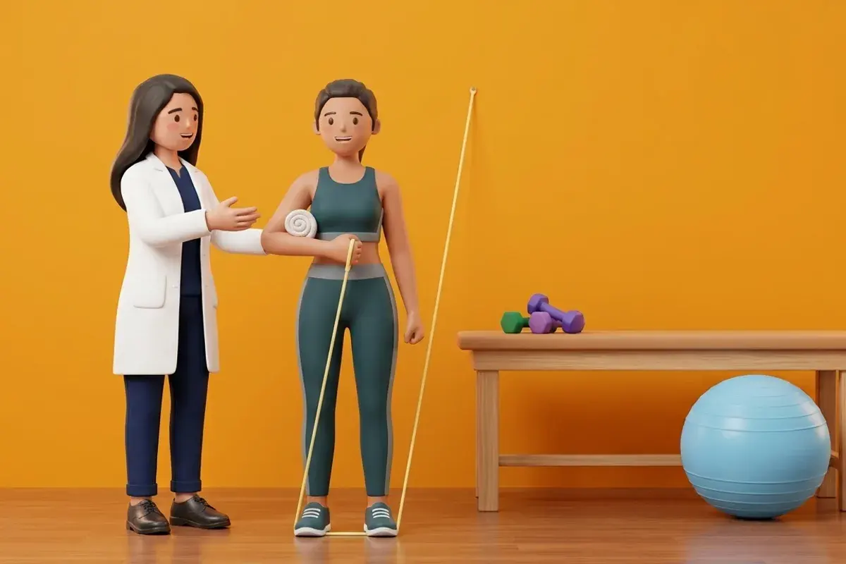
- Progressive FCU Isometric and Concentric Strengthening (Weeks 4 to 8): Initiate submaximal isometric wrist flexion and ulnar deviation against resistance bands; progress to dynamic eccentric wrist curling with light dumbbells to restore powerful grip and racket stability.
Frequently Asked Questions
What is the pisiform bone, and why does pushing on the heel of my palm hurt?
The pisiform is a small, pea-sized bone embedded right inside your main wrist-bending muscle tendon (the FCU), like a kneecap inside your knee. When you fall hard on your palm or play sports with heavy impact, the cartilage under this tiny bone wears down, causing sharp pain whenever you lean on your palm.
If the surgeon takes out my pisiform bone, will my wrist become weak?
No! Because the surgeon carefully shells the bone out from inside the tendon while leaving the tendon itself completely attached to your hand, your wrist retains full bending power and strength, while the bone-on-bone grinding pain is permanently gone.
How can my therapist tell if my wrist pain is a TFCC tear or pisiform arthritis?
In the pisotriquetral grind test, your therapist holds your wrist completely relaxed and wiggles the small pea-bone back and forth with their fingers. If this reproduces your exact pain and creates a rough crunching sound, the problem is in your pisiform joint, not the deeper TFCC cartilage.
Related condition guides

Related recovery guides & articles
BookPhysio.in Editorial Team
The BookPhysio.in editorial team writes and maintains this content, checking it against reliable clinical sources. Content follows our medical review policy: general information only, and no page claims a clinical review until a named registered physiotherapist has signed it off.
Read our medical review policy