
Plantar Fasciitis versus Fasciopathy: Histopathology, Collagen Breakdown, and Tensile Strain
In short
Understand the true histopathological transition from acute plantar inflammation to chronic degenerative fasciopathy, collagen breakdown, and restorative rehabilitation.
In this guide (4 sections)
Plantar heel pain has historically been termed plantar fasciitis under the clinical assumption that persistent symptoms originate from chronic acute inflammation of the plantar aponeurosis. However, extensive histological, biochemical, and sonographic evaluations over the past two decades have demonstrated that the condition is fundamentally a degenerative fasciopathy rather than an active inflammatory disorder. Surgical biopsy specimens harvested from individuals with recalcitrant inferior heel pain consistently demonstrate an absence of classical inflammatory cell infiltrates, such as neutrophils, macrophages, or lymphocytes. Instead, tissue analyses reveal non-inflammatory angiofibroblastic hyperplasia, marked hypervascularity with disorganized capillary buds, extracellular matrix breakdown, and fragmentation of type I collagen bundles replaced by weak, immature type III collagen. Recognizing this condition as plantar fasciopathy completely reframes the rehabilitation framework, shifting intervention from passive anti-inflammatory modalities toward targeted mechanical tensile loading that drives cellular collagen remodeling.
Barefoot Marble Floor Living, Prolonged Kitchen Standing, and Thin Flat Slippers
In Indian households, domestic surface architecture and cultural footwear habits play a profound role in propagating chronic tensile strain across the plantar fascia. The vast majority of residences in urban cities such as Mumbai, Delhi, Bengaluru, and Chennai feature unyielding polished marble, vitrified ceramic tile, or granite flooring. Walking completely barefoot across these stiff surfaces deprives the heel of natural impact attenuation, transferring raw ground reaction forces directly into the medial calcaneal enthesis during every heel strike. Furthermore, homemakers frequently spend three to five continuous hours standing barefoot or wearing paper-thin flat rubber chappals while preparing meals at elevated kitchen counters. These flat slippers provide zero longitudinal arch support and no heel elevation, forcing the plantar aponeurosis to remain under maximal passive tensile strain. When combined with long commute walks over uneven concrete pavements or prolonged market standing, the mechanical load rapidly exceeds the biological healing capacity of the enthesis.
Windlass Mechanism Strain, Calcaneal Enthesopathy, and Plantar Thickness
The plantar aponeurosis is a dense fibrous band originating primarily from the medial process of the calcaneal tuberosity and fanning out distally to insert into the plantar plates and bases of the proximal phalanges. Under weight-bearing conditions, the fascia acts as a primary passive tie-rod for the medial longitudinal arch. During the terminal stance and pre-swing phases of gait, extension of the metatarsophalangeal joints winds the plantar fascia around the metatarsal heads, shortening the distance between the calcaneus and metatarsals while elevating the arch, a mechanical phenomenon known as the Windlass mechanism. In chronic fasciopathy, repetitive micro-tearing occurs at the calcaneal insertion where tensile stresses are highest. The tissue responds with excessive proteoglycan accumulation, chondroid metaplasia of native fibroblasts, and loss of normal parallel fiber alignment. High-resolution diagnostic ultrasound characteristically demonstrates marked pathological thickening of the proximal fascia exceeding the standard four-millimeter threshold, accompanied by loss of crisp fibrillar architecture and localized hypoechogenicity.
Rathleff High-Load Towel Training, Low-Dye Taping, and Intrinsic Foot Doming
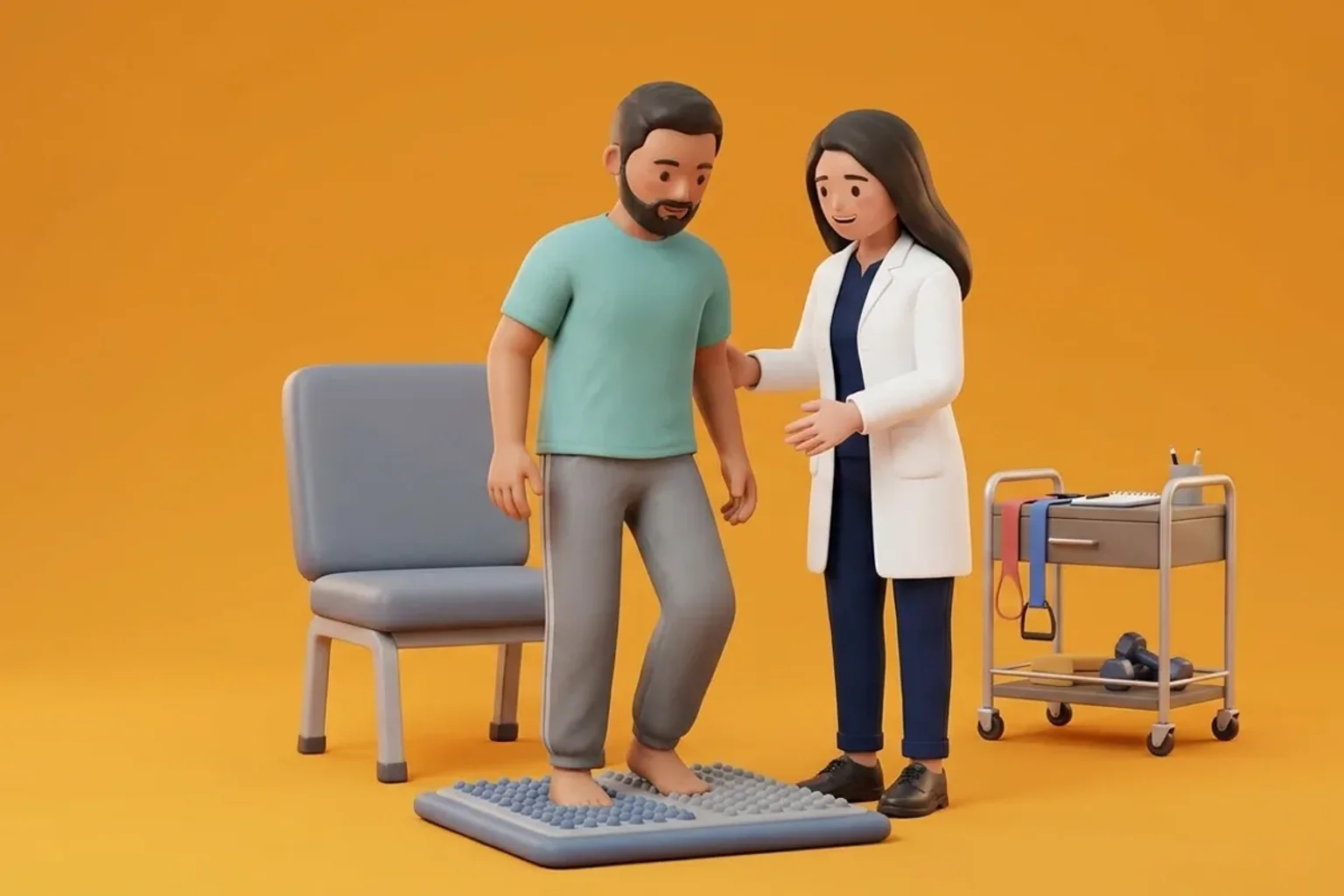
Because plantar fasciopathy is characterized by cellular degeneration and failed matrix adaptation rather than acute inflammatory exudation, passive rest and long-term anti-inflammatory medications provide limited durable benefit. Contemporary physiotherapy centers upon progressive tensile loading to stimulate tenocyte and fibroblastic mechanotransduction, promoting the synthesis of organized type I collagen. The landmark Rathleff protocol utilizes high-load, slow eccentric-concentric calf raises with a rolled towel placed beneath the toes to maximally engage the Windlass mechanism. This specific positioning pre-tensions the plantar fascia while the gastrocnemius-soleus complex contracts, delivering controlled tensile stress that encourages structural fiber realignment. Concurrently, therapeutic low-Dye taping provides immediate mechanical unloading of the longitudinal arch during acute exacerbations, reducing morning first-step pain. Intrinsic foot muscle training, particularly short-foot doming drills, strengthens the active muscular arch supports, effectively shielding the healing fascial band from excessive tensile deformation.
Rathleff High-Load Heel Raise with Great Toe Towel Windlass Extension
Objective: Deliver controlled tensile mechanical strain to the plantar fascia to stimulate collagen remodeling and cellular mechanotransduction
- 1Place a sturdy aerobic step or stair edge near a stable wall or handrail for balance support.
- 2Roll a small hand towel tightly into a firm cylinder approximately one to two inches in diameter and place it across the step edge.
- 3Position your foot so that your metatarsal heads rest firmly on the step surface while your toes extend upward over the towel roll, maximally engaging the Windlass mechanism.
- 4Over a slow count of 3 seconds, rise upward into a full single-leg heel raise, feeling your calf contract and tensile tension engage your foot arch.
- 5Pause at the top for a full 2-second isometric contraction, then slowly lower your heel below the step level over a controlled 3-second descent.
| Diagnostic and Pathological Domain | Acute Plantar Fasciitis (Initial 2-4 Weeks) | Chronic Plantar Fasciopathy (Beyond 6-12 Weeks) | Targeted Physiotherapy Strategy |
|---|---|---|---|
| Histological Profile | Perifascial edema, occasional micro-vascular leakage, initial inflammatory response | Non-inflammatory angiofibroblastic hyperplasia, matrix disorganization, collagen breakdown | Shift from passive anti-inflammatory rest to tensile mechanotherapy and loading |
| Ultrasound Characteristics | Mild perifascial fluid halo with preserved internal fibrillar striations | Fusiform thickening exceeding 4.0 mm, internal hypoechogenicity, loss of fibrillar pattern | Serial sonographic monitoring of thickness during progressive strength loading |
| Response to Rest vs Loading | Temporary symptom relief with complete unloading and ice application | Persistent stiffness upon first steps after rest; gradual warming with light motion | High-load slow resistance training (Rathleff protocol) and intrinsic arch doming |
While plantar heel pain is predominantly caused by chronic fasciopathy, clinicians must remain alert for red flag conditions. A sudden, audible pop under the arch during running or jumping followed by immediate ecchymosis and severe weight-bearing inability suggests an acute fascial rupture. Severe pain elicited by bilateral medial-lateral compression of the posterior calcaneal body (positive squeeze test) strongly indicates a calcaneal stress fracture. Furthermore, persistent burning or radiating paresthesia along the lateral plantar border suggests Baxter nerve entrapment. In such presentations, obtain immediate orthopaedic evaluation and diagnostic imaging.
Frequently Asked Questions
Why does my heel hurt worst during the first steps out of bed in the morning?
During sleep, your foot naturally rests in an unloaded plantarflexed and slightly inverted posture. In this shortened position, the plantar fascia cools, tightens, and attempts to repair microscopic collagen tears with immature, fragile cross-links. When you step out of bed and bear full body weight on hard flooring, the fascia is abruptly stretched under intense tensile load, re-tearing those delicate cross-links and irritating sensitized nociceptors. Performing seated great-toe extension stretches and gentle calf warm-ups before your feet touch the floor prevents this abrupt morning strain.
Do I need a doctor referral to treat plantar fasciitis on BookPhysio.in?
BookPhysio.in does not require a doctor referral. Under the National Commission for Allied and Healthcare Professions (NCAHP) Act 2021, patients across India enjoy direct access to autonomous registered physiotherapists. Through BookPhysio.in, you connect directly with verified clinicians and pay per session with transparent fees, completely free from advance package commitments, commission markups, or platform subscription charges.
Related condition guides
Related recovery guides & articles
BookPhysio.in Editorial Team
The BookPhysio.in editorial team writes and maintains this content, checking it against reliable clinical sources. Content follows our medical review policy: general information only, and no page claims a clinical review until a named registered physiotherapist has signed it off.
Read our medical review policy