
Post-Operative Meniscal Repair Rehab: Six-Week Flexion Restriction and Protection Protocol
In short
Master the biological rationale and clinical milestones of the critical six-week post-operative protection phase following meniscal repair surgery.
In this guide (4 sections)
Arthroscopic meniscal repair represents the gold-standard surgical technique for preserving native fibrocartilage and protecting long-term knee biomechanics in patients with repairable tear patterns. However, the success of any meniscal repair is fundamentally dependent on rigorous adherence to post-operative biological protection protocols during the first six weeks. Surgical implants, whether all-inside suture anchors, outside-in stitches, or inside-out horizontal mattresses, merely approximate the torn fibrocartilaginous margins under initial mechanical tension. True biological healing requires months of cellular migration, microvascular angiogenesis, fibrocartilage matrix synthesis, and collagen cross-linking. During the initial six-week vulnerable window, unconstrained knee flexion or premature rotational shear can readily pull sutures through fragile meniscal tissue, leading to silent repair failure and joint space deterioration.
Navigating Indian Domestic Recovery: Western Commode Adjustments and Kitchen Roles
Managing the first six weeks following meniscal repair in an Indian home requires strict family coordination and domestic modification. Indian household traditions of gathering on living room floor carpets, praying in deep kneeling positions, and cooking near floor level must be temporarily paused. The patient must wear a rigid hinged knee brace locked in extension for ambulation and use axillary or elbow crutches for protected partial weight-bearing. In homes with Indian-style squatting toilets, installing a portable commode chair over the pan is essential before discharge. Household family members must be educated that asking the patient to bend their knee past 90 degrees to sit in low auto-rickshaws or floor chatais risks tearing out surgical sutures. Organizing a ground-floor bedroom with a high bed and clear walking pathways prevents accidental falls on polished tile floors.
Biomechanics of Suture Strain: The 90-Degree Flexion Threshold
The universal clinical mandate restricting knee flexion to no more than 90 degrees during the initial six weeks is grounded in precise joint kinematics. Between 0 and 60 degrees of knee flexion, the femoral condyles roll primarily over the anterior and middle meniscal body, maintaining low shear forces across posterior horn repairs. However, as the knee flexes past 90 degrees toward deep flexion, the femoral condyles slide backward onto the posterior meniscal horns. This backward excursion applies powerful posterior shear and hoop stress directly against the healing repair site. Biomechanical cadaveric studies demonstrate that suture displacement forces rise exponentially beyond 90 degrees of flexion, frequently exceeding the 30 to 100 Newton pullout limit of meniscal repair devices. Limiting flexion strictly to 90 degrees protects the delicate fibrovascular bridging tissue from mechanical failure.
Structured Phase I Milestones: Patellar Mobility, Isometric Quads, and Passive Range
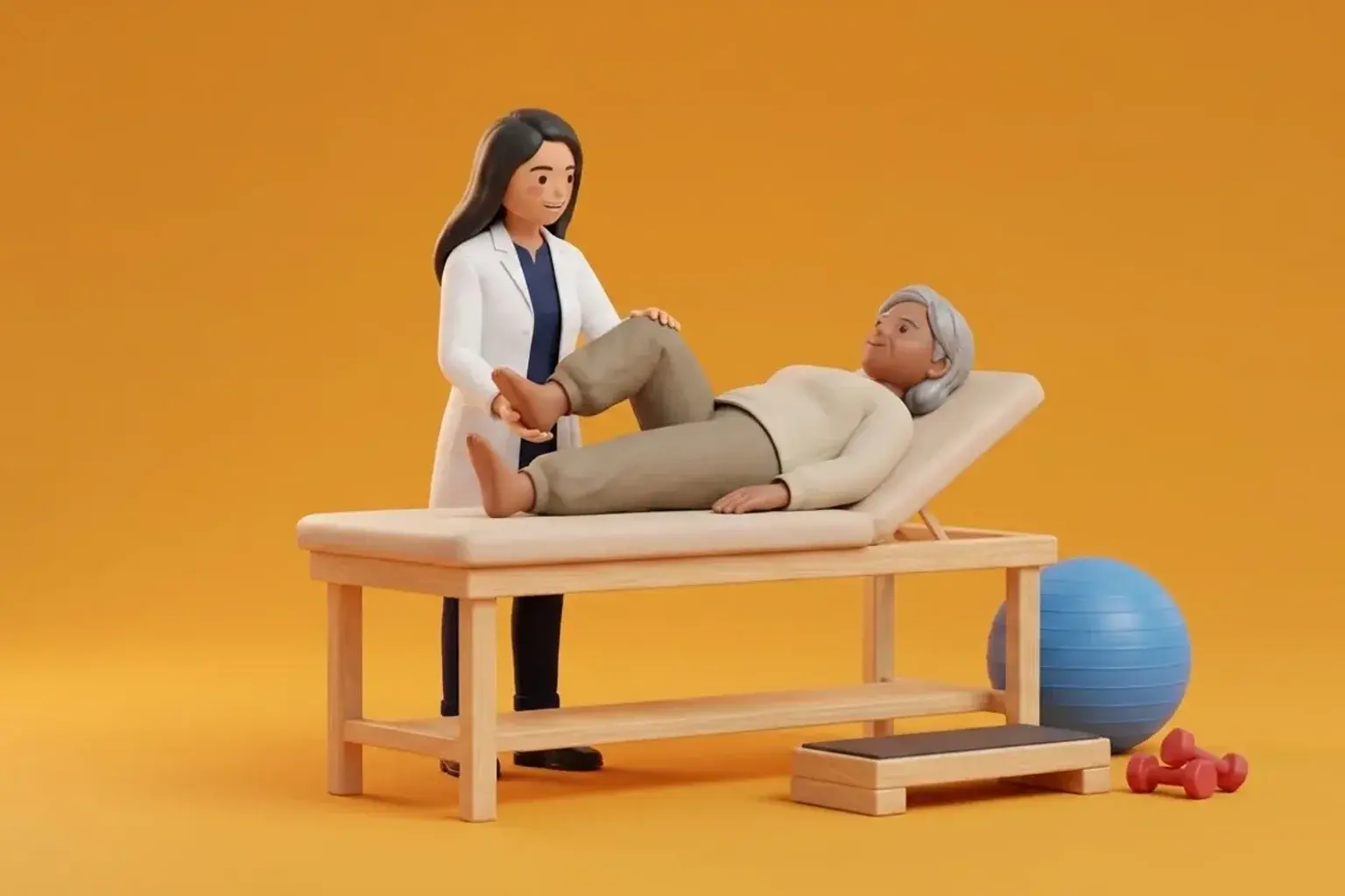
Phase I rehabilitation (Weeks 0 to 6) centres on four foundational objectives: protecting the surgical repair, preventing patellofemoral adhesions, restoring full passive extension, and overcoming arthrogenic quadriceps inhibition. The hinged knee brace is locked at 0 degrees for ambulation with crutches, allowing 25 to 50 percent partial flat-foot weight-bearing. Dedicated manual therapy includes gentle superior, inferior, and medial patellar mobilizations performed twice daily to prevent infrapatellar fat pad contracture. Range of motion exercises are strictly performed in a passive or active-assisted manner between 0 and 90 degrees in non-weight-bearing positions (such as seated plinth slides or supine wall slides). Strengthening during this phase is confined strictly to non-shear drills, including quadriceps sets, straight leg raises in all four planes with the brace locked, and seated ankle pumps.
Supine Wall Slide with 90-Degree Stop Marker
Objective: Regain controlled passive-assisted knee flexion up to exactly 90 degrees without placing shear force on the repaired meniscus
- 1Lie comfortably flat on your back on an exercise mat with your buttocks positioned approximately 30 to 40 centimetres from a smooth wall.
- 2Place the heel of your surgical leg against the wall with your knee straight and your foot wearing a smooth sock.
- 3Place the foot of your non-surgical, unaffected leg underneath the heel of your surgical leg to act as a supportive brake.
- 4Slowly allow the surgical heel to slide down the wall under gravity, using your opposite foot to control the descent speed.
- 5Stop the slide immediately when the knee reaches a right angle (90 degrees), pause for 5 seconds, and use the opposite leg to push the heel back up to straight.
| Rehabilitation Component | Weeks 0 to 2 Guidelines | Weeks 3 to 4 Guidelines | Weeks 5 to 6 Guidelines |
|---|---|---|---|
| Weight-Bearing Status | Toe-touch to 25% weight-bearing with crutches | 50% partial weight-bearing with crutches | 75% to full weight-bearing as tolerated in locked brace |
| Hinged Knee Brace | Locked at 0 deg for all walking and sleep | Locked at 0 deg for walking; unlocked 0-90 for sitting | Unlocked 0-90 deg for walking if quad control is excellent |
| Permitted Flexion Range | Strictly 0 to 60 degrees passive/assisted | Progressed smoothly from 0 to 75 degrees | Progressed to a strict maximum of 90 degrees |
| Core Exercise Drills | Quad sets, patellar glides, ankle pumps | Braced straight leg raises, prone terminal extensions | Seated hamstring curls (0-60 deg), standing hip abduction |
While protecting against excessive flexion is critical, failing to achieve full passive knee extension (0 degrees) within the first 2 to 3 weeks leads to permanent flexion contractures, patellofemoral pain, and abnormal gait kinetics. If a rubbery mechanical block prevents complete extension despite regular quad sets and prone hangs, the clinician must assess for arthrofibrosis or anterior interval scar tethering, alerting the orthopaedic surgical team.
Frequently Asked Questions
Why does my knee feel tight and stiff if I can only bend it to 90 degrees for six weeks?
Temporary joint stiffness is an expected and necessary trade-off to protect your healing meniscus sutures from tearing. Restricting flexion to 90 degrees ensures the repair site heals firmly without mechanical disruption. Once the initial six-week biological consolidation window passes, your physiotherapist will safely guide you through progressive stretching to regain your full natural knee flexion.
Do I need a doctor referral to treat a meniscal tear on BookPhysio.in?
BookPhysio.in does not require a doctor referral. Under the National Commission for Allied and Healthcare Professions (NCAHP) Act 2021, patients across India enjoy direct access to autonomous registered physiotherapists. Through BookPhysio.in, you connect directly with verified clinicians and pay per session with transparent fees, completely free from advance package commitments, commission markups, or platform subscription charges.
Related condition guides



Related recovery guides & articles
BookPhysio.in Editorial Team
The BookPhysio.in editorial team writes and maintains this content, checking it against reliable clinical sources. Content follows our medical review policy: general information only, and no page claims a clinical review until a named registered physiotherapist has signed it off.
Read our medical review policy