
Thessaly Test at Five and Twenty Degrees: Dynamic Rotational Meniscal Stress Mechanics
In short
Examine the dynamic weight-bearing rotational mechanics of the Thessaly test at 5 and 20 degrees, clinical execution, and diagnostic advantages.
In this guide (4 sections)
Introduced by Karachalios and colleagues in 2005, the Thessaly test is a dynamic, weight-bearing physical examination maneuver designed to reproduce the physiological stresses that provoke meniscal symptoms during daily functional activities. Unlike traditional non-weight-bearing tests such as the McMurray or Apley maneuvers, which apply passive torque to an unloaded limb, the Thessaly test combines axial body weight compression with active internal and external rotation of the femur over the fixed tibia. The test is performed sequentially at two distinct angles of knee flexion: first at 5 degrees to establish a baseline and evaluate peripheral stability, and subsequently at 20 degrees to subject the meniscal bodies and posterior horns to maximal compressive shear. Biomechanical analyses confirm that weight-bearing rotation transmits higher hoop and shear stresses across fibrocartilage, significantly enhancing diagnostic sensitivity for subtle tears.
Navigating Indian Domestic Pivoting: Narrow Hallways and Scooter Mounting
The dynamic mechanics of the Thessaly test mirror frequent rotational challenges encountered in Indian living environments. When an individual steps into a compact bathroom, pivots around narrow doorways, or turns while holding a hot cooking pan in a modest kitchen, the stance knee undergoes axial rotation under full body mass. Similarly, mounting a two-wheeler requires placing one foot firmly on the pavement while swinging the opposite leg across the seat, subjecting the weight-bearing knee to 15 to 25 degrees of flexion paired with sharp rotational torque. When a patient reports that their knee catches or feels unstable during these everyday domestic pivots, reproducing the exact mechanical vector via the Thessaly test helps the physiotherapist confirm whether meniscal shear is the underlying trigger, guiding functional movement retraining.
Tibiofemoral Contact Vectors, Posterior Horn Excursion, and Torsional Shearing
The diagnostic efficacy of the Thessaly test stems from joint contact mechanics. At 5 degrees of knee flexion, the tibiofemoral contact area is located primarily over the anterior and middle portions of the menisci, and axial rotation produces relatively modest shear forces. When the knee flexes to 20 degrees, the femoral condyles roll posteriorly, shifting peak compressive contact directly onto the broader, thicker posterior meniscal horns. Internal rotation of the femur on the fixed tibia translates the medial femoral condyle posteriorly, squeezing the medial meniscus posterior horn against the posterior tibial rim. External rotation of the femur similarly compresses the lateral meniscus. If an unstable longitudinal flap, radial tear, or horizontal cleavage split is present, this torsional compression jams the torn fragment, eliciting localized joint line discomfort, mechanical clicking, or a sensation of the knee giving way.
Execution Protocol, Safety Precautions, and Proprioceptive Retraining
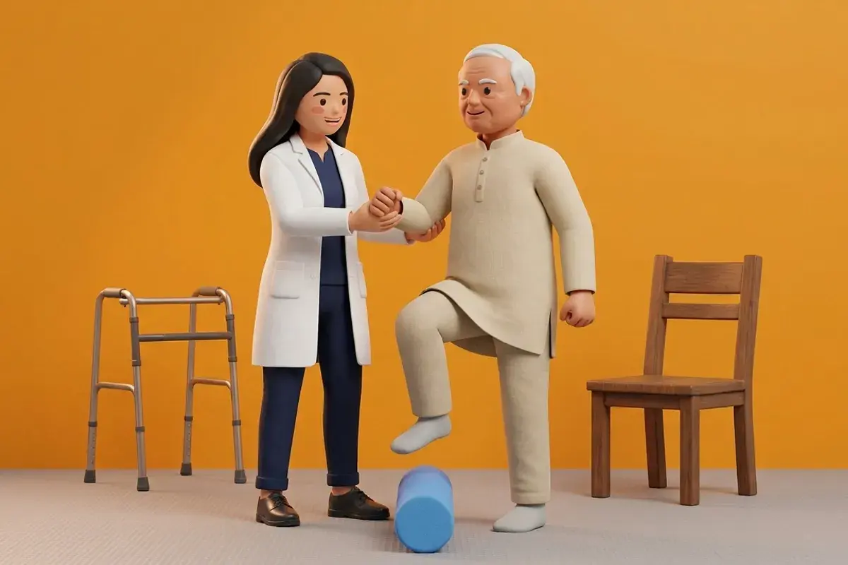
To execute the test accurately, the patient stands on the affected single limb while holding the examiner outstretched hands for balance. The patient flexes the knee to 20 degrees and actively rotates the trunk and pelvis internally and externally three times in each direction, keeping the foot planted firmly on the floor. A positive test requires reproduction of familiar joint line pain, mechanical catching, or locking. Because the test introduces substantial rotational torque, it is contraindicated in patients with acute knee effusion, severe resting pain, or suspected gross anterior cruciate ligament tears. Following testing, rehabilitation addresses identified rotational deficits through closed-chain hip external rotator strengthening, ankle stability drills, and multiplanar balance training to protect the meniscus during daily pivoting.
Supported Single-Leg Clock Reaches for Closed-Chain Stability
Objective: Enhance multiplanar knee alignment and hip abductor control during controlled weight-bearing pivot movements
- 1Stand tall on your affected leg next to a sturdy dining table or kitchen counter, resting light fingertips for balance.
- 2Unlock your stance knee into approximately 15 to 20 degrees of soft flexion, keeping your kneecap aligned over your second toe.
- 3Keeping your weight firmly centered over the stance foot, reach your opposite foot forward to touch the imaginary 12 o clock spot on the floor.
- 4Return to center, then reach outward to the 3 o clock spot (or 9 o clock for left leg), maintaining steady knee stability.
- 5Finally, reach backward to the 6 o clock spot, ensuring the stance knee does not collapse inward into excessive valgus.
| Test Angle | Biomechanical Focus | Sensitivity Range | Specificity Range | Clinical Indication |
|---|---|---|---|---|
| 5 Degrees Knee Flexion | Anterior and middle meniscal body loading; low shear baseline | 55% to 65% | 60% to 75% | Initial screening; ruling out severe irritability before 20-degree test |
| 20 Degrees Knee Flexion | Posterior horn compression and maximal rotational shear | 75% to 90% | 70% to 85% | Primary provocative assessment for meniscal tears and root pathology |
| Concomitant ACL Tear | Combined tibiofemoral subluxation and rotational instability | Reduced diagnostic accuracy | High false-positive rate | Avoid or interpret with extreme caution due to joint instability |
The Thessaly test should never be performed on an acutely injured knee with a tense effusion, gross hemarthrosis, or significant ligamentous laxity (such as a Grade III ACL or MCL tear). Applying axial rotational torque to an unstable, unconstrained knee can provoke subluxation, damage secondary stabilizing chondral surfaces, or cause severe acute distress. Clinicians must clear ligamentous integrity and acute inflammation before conducting weight-bearing rotational tests.
Frequently Asked Questions
Is the Thessaly test better than an MRI for finding a meniscal tear?
The Thessaly test is an excellent dynamic clinical screening tool that helps your physiotherapist determine if your pain is reproduced by meniscal compression. However, it does not replace magnetic resonance imaging. While the Thessaly test demonstrates high sensitivity for detecting symptomatic tears, MRI remains necessary to evaluate exact tear depth, vascular zone, root involvement, and underlying cartilage integrity.
Do I need a doctor referral to treat a meniscal tear on BookPhysio.in?
BookPhysio.in does not require a doctor referral. Under the National Commission for Allied and Healthcare Professions (NCAHP) Act 2021, patients across India enjoy direct access to autonomous registered physiotherapists. Through BookPhysio.in, you connect directly with verified clinicians and pay per session with transparent fees, completely free from advance package commitments, commission markups, or platform subscription charges.
Related condition guides



Related recovery guides & articles
BookPhysio.in Editorial Team
The BookPhysio.in editorial team writes and maintains this content, checking it against reliable clinical sources. Content follows our medical review policy: general information only, and no page claims a clinical review until a named registered physiotherapist has signed it off.
Read our medical review policy