
Tibial Plateau Fracture Rehabilitation: Schatzker Types I to VI Knee Mobilisation
In short
Tibial plateau articular fractures require delicate balancing between early range of motion to prevent arthrofibrosis and strict joint offloading to protect articular cartilage. Discover clinical protocols across Schatzker classifications.
In this guide (6 sections)
Tibial plateau fractures disrupt the weight-bearing articular surface of the proximal tibia, frequently accompanied by meniscal detachment and cruciate ligament compromise. The Schatzker classification categorizes these injuries from Type I (cleave split of the lateral plateau) through Types II and III (split-depression and central depression), Type IV (high-energy medial plateau split), Type V (bicondylar fracture), to Type VI (metaphyseal-diaphyseal dissociation). Surgical management with anatomical articular reduction, subchondral bone grafting, and rigid locking plates aims to restore a congruent joint surface. Post-operative physiotherapy must navigate a tight biological window: initiating early passive motion to prevent devastating knee stiffness while enforcing strict non-weight bearing to prevent articular step-off and secondary osteoarthritis.
Domestic and Cultural Indian Realities
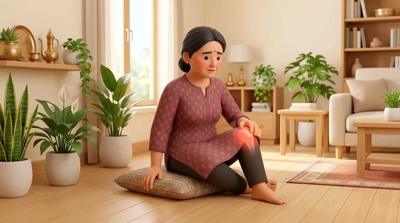
Indian domestic life involves substantial knee flexion during everyday tasks. Deep squatting for Indian commodes places contact forces up to seven times body weight across the tibial plateaus, which will crush healing subchondral bone graft; installing a raised toilet seat chair over the squat pan is an absolute necessity. Deep floor sitting for family meals and pooja rituals must be replaced by dining at standard table heights and using an elevated prayer chowki that keeps the operated knee in a comfortable 45 to 60 degree position. Low rope charpoys must be elevated using hardwood bed risers to establish a 20-inch transfer height, preventing the patient from forcing the knee into acute flexion while standing up. When commuting, travelling in shared autorickshaws or two-wheelers creates severe torsional and axial jolts; patients must use air-conditioned cars or non-emergency medical cabs with the leg resting extended on the rear seat during the initial 8 weeks.
Clinical Biomechanics and Anatomical Healing
The tibial plateau bears axial compressive loads transmitted from the femoral condyles. When articular cartilage and subchondral bone undergo structural reduction, subchondral rafting screws hold the plateau elevation. Loading the limb prematurely produces compressive shear that can cause hardware loosening and articular collapse greater than 2mm, directly precipitating post-traumatic arthrosis. However, complete joint immobilisation causes synovial adhesion, capsular shortening, and quadriceps arthrogenic muscle inhibition. Continuous passive motion (CPM) or active-assisted heel slides within a controlled arc (0 to 90 degrees) nourishes articular chondrocytes through synovial fluid diffusion without generating axial contact pressure.
Validated Special Tests and Differential Diagnosis
Post-operative assessment monitors patellar mobility via superior-inferior and mediolateral patellar glides, which are essential prerequisites for normal knee flexion. Ligamentous stability tests (Lachman, pivot shift, varus and valgus stress tests) are deferred until bony union is secured to protect repaired capsular structures. Neurovascular evaluation focuses on the common peroneal nerve, which wraps around the fibular neck and is vulnerable in lateral split-depression fractures (Schatzker I to III), manifesting as numbness in the first dorsal web space and weakness in foot eversion and dorsiflexion.
Stage-by-Stage Functional Recovery Protocol
During Weeks 0 to 2, the knee is maintained in a hinged knee brace locked at 0 degrees for transfers and sleep, unlocking for passive and active-assisted range of motion from 0 to 60 degrees. Patients perform static quadriceps sets, patellar mobilisations, and active ankle pumps. From Weeks 2 to 6, flexion is gradually increased to 90 degrees, incorporating seated active-assisted knee extensions and straight leg raises with the brace locked. Weeks 6 to 12 mark the introduction of progressive partial weight-bearing (starting at 25% body weight with a bathroom scale guide) once serial X-rays confirm bridging bone, advancing to full weight-bearing and closed-chain mini-squats by week 12.
Seated Active-Assisted Heel Slides with Towel
Objective: Restore knee flexion safely without compressive axial joint loading
- 1Sit upright on a firm wooden chair with feet resting on a smooth tiled or wooden floor.
- 2Loop a long cotton towel or yoga strap around the sole of the foot on the operated leg.
- 3Gently slide the heel backward toward the chair by pulling gently on the strap handles while actively contracting the hamstring muscles.
- 4Pause at the point of gentle stretch for 5 seconds without forcing into sharp pain.
- 5Slowly slide the heel forward back to the starting resting position using quadriceps control.
| Schatzker Type | Articular Complexity | Early Motion Arc (Weeks 0 to 6) | Weight-Bearing Progression |
|---|---|---|---|
| Type I to III (Lateral Split/Depression) | Partial articular involvement | 0 to 90 degrees active-assisted | Non-weight bearing 6 weeks; partial 6 to 10 weeks; full by 12 weeks |
| Type IV (Medial Plateau) | High-energy medial shear | 0 to 60 degrees first 3 weeks, then 90 | Strict non-weight bearing 8 to 10 weeks due to varus instability |
| Type V & VI (Bicondylar / Dissociation) | Complete articular disruption | 0 to 45 degrees early CPM, gradual to 90 | Strict non-weight bearing 10 to 12 weeks until solid metaphyseal callus |
Red Flags and Urgent Surgical Indicators
Immediate surgical consultation is warranted if the patient experiences severe tense calf swelling with excruciating pain on passive toe stretch, indicating acute compartment syndrome. Other urgent signs include sudden loss of active ankle dorsiflexion (peroneal nerve compromise), wound dehiscence with persistent purulent exudate, or an acute localized sensation of structural clicking accompanied by sudden angular deformity, signaling hardware breakage or plateau collapse.
Frequently Asked Questions
Can I use continuous passive motion (CPM) machines at home in India?
Yes, CPM machines can be rented for domiciliary use under your physiotherapist direction. They assist in gently maintaining knee joint mobility during the early non-weight bearing weeks, particularly for complex Schatzker types V and VI, preventing joint stiffness while keeping axial joint stresses minimal.
When can I begin walking without crutches after plateau surgery?
Independent unassisted walking is generally cleared only after 12 to 14 weeks, once follow-up orthopaedic radiographs demonstrate robust trabecular bridging and no articular step-off. Progressing from two crutches to a single stick, and finally unassisted gait, must be clinically supervised to prevent persistent limp and joint overload.
Related condition guides



Related recovery guides & articles
BookPhysio.in Editorial Team
The BookPhysio.in editorial team writes and maintains this content, checking it against reliable clinical sources. Content follows our medical review policy: general information only, and no page claims a clinical review until a named registered physiotherapist has signed it off.
Read our medical review policy