
Unilateral Vestibular Hypofunction: Vestibular Neuritis Recovery
In short
Vestibular neuritis produces sudden unilateral vestibular hypofunction. Learn how targeted VOR adaptation exercises accelerate central compensation.
In this guide (6 sections)
Unilateral Vestibular Hypofunction (UVH) most commonly arises following acute vestibular neuritis or labyrinthitis, typically triggered by reactivation of herpes simplex virus type 1 affecting the superior vestibular nerve. The sudden, unilateral loss of tonic resting vestibular afferent discharge (normally 70 to 100 spikes per second) creates a massive firing asymmetry within the brainstem vestibular nuclei. This physiological imbalance produces an acute vestibular crisis: severe rotational vertigo, vegetative autonomic distress, spontaneous horizontal-torsional nystagmus beating away from the damaged ear, and profound postural deviation toward the side of the lesion.
Indian Domestic Realities and Bathroom Slip Prevention
During the acute and subacute recovery phases of vestibular neuritis, patients in Indian households face high fall risks during early morning routines. Navigating wet, smooth tiled bathrooms to fetch water buckets (baltis) or stepping over raised stone door thresholds (dehleez) in dim light frequently induces severe unsteadiness. Family members often encourage the patient to remain in a darkened room resting completely, but prolonged immobility delays central cerebellar compensation. Physiotherapists intervene early, installing non-slip bathroom mats, improving hallway night illumination, and initiating active head movement protocols.
Static vs Dynamic Cerebellar Compensation Mechanisms
Recovery from UVH occurs in two distinct physiological stages. Static compensation resolves spontaneous nystagmus and resting postural tilt within 3 to 7 days through neurochemical rebalancing of gamma-aminobutyric acid (GABA) and glutamate receptors within the deafferented medial vestibular nucleus. In contrast, dynamic compensation, which restores the vestibulo-ocular reflex (VOR) gain during rapid head movement, never occurs spontaneously with rest. Dynamic compensation requires active retinal slip, where visual error signals drive neuroplastic synaptic remodeling in the cerebellar flocculus and vestibular nuclei.
Apply the three-step HINTS examination (Head Impulse, Nystagmus, Test of Skew). A normal head impulse test, direction-changing gaze-evoked nystagmus, or vertical skew deviation indicates an acute posterior circulation stroke requiring immediate emergency neurovascular intervention.
Evidence-Based Vestibular Rehabilitation Protocol
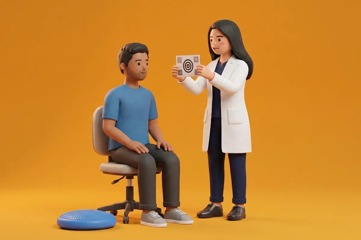
Vestibular rehabilitation therapy must commence as soon as severe nausea is controlled, ideally within 72 to 96 hours of symptom onset. Vestibular suppressants like dimenhydrinate or promethazine must be weaned immediately, as they directly block cerebellar compensation. The patient performs structured VOR adaptation drills (VOR x1 and VOR x2) at varying speeds and distances, combined with dynamic balance retraining on compliant surfaces.
Seated VOR x1 Horizontal Gaze Stabilization Exercise
Objective: Stimulate cerebellar synaptic plasticity and restore vestibulo-ocular reflex gain
- 1Sit upright on a supportive chair facing a blank wall 1 meter away.
- 2Hold a small target with an uppercase letter printed on it at eye level directly in front of your face.
- 3Fix both eyes firmly on the target letter, keeping it sharply in focus.
- 4Rotate your head horizontally side-to-side 20 degrees in each direction at a speed of 1 to 2 cycles per second.
- 5Ensure the letter remains perfectly clear and still throughout the movement without blurring or jumping.
Stages of Vestibular Compensation in Unilateral Hypofunction
| Compensation Stage | Timeframe | Underlying Neurobiology | Clinical Presentation | Primary Physiotherapy Focus |
|---|---|---|---|---|
| Acute Static Stage | Days 1 to 7 | GABAergic receptor recalibration in vestibular nuclei | Spontaneous nystagmus in primary gaze; severe nausea | Early gentle mobilization; wean vestibular suppressants |
| Subacute Dynamic Stage | Weeks 2 to 6 | Cerebellar long-term depression driven by retinal slip | Gaze instability (oscillopsia) during rapid head turns | VOR x1 adaptation and dynamic weight shifts |
| Chronic Consolidation | Months 2 to 6 | Consolidated neuroplastic pathways and sensory re-weighting | Occasional unsteadiness in dark or complex environments | Dual-tasking, compliant foam balance, community walking |
Red Flags Demanding Emergency Stroke Workup
Immediate hospital transfer for stroke protocol is required if dizziness presents with a negative head impulse test, direction-changing nystagmus, vertical ocular skew, limb weakness, or acute hearing loss in a patient with cardiovascular risk factors.
Frequently Asked Questions
Will my inner ear nerve completely regenerate after vestibular neuritis?
In many cases, peripheral nerve damage is partial and some inner ear function returns. However, full functional recovery does not depend on nerve regeneration; it relies on your brain learning to compensate and recalibrate its balance circuits through active physiotherapy exercises.
Can I book an expert vestibular physiotherapist through BookPhysio.in?
Yes. BookPhysio.in connects you directly with verified neurological and vestibular physiotherapists across India. Under the NCAHP Act 2021, you do not require a hospital referral, and you pay transparently per session without advance package locks.
Related condition guides

Related recovery guides & articles
BookPhysio.in Editorial Team
The BookPhysio.in editorial team writes and maintains this content, checking it against reliable clinical sources. Content follows our medical review policy: general information only, and no page claims a clinical review until a named registered physiotherapist has signed it off.
Read our medical review policy