
Vestibular Neuritis: Acute Phase Management, Spontaneous Nystagmus and Early Rehabilitation
In short
Acute vestibular neuritis causes sudden severe rotational vertigo without hearing loss; early cessation of vestibular suppressants and timely rehabilitation promote central compensation.
In this guide (8 sections)
Pathophysiology of Acute Vestibular Deafferentation
Vestibular neuritis is an acute, unilateral peripheral vestibular disorder caused by inflammation of the vestibular nerve, most commonly the superior division. Believed to result from viral infection or reactivation of herpes simplex virus type 1 (HSV-1) within Scarpa's ganglion, it triggers sudden unilateral deafferentation. The resting firing rate of vestibular afferents from the affected labyrinth collapses from its baseline of approximately 90 spikes per second toward zero.
This profound electrical imbalance between the vestibular nuclei is interpreted by the brainstem as a rapid, violent head rotation toward the intact ear. Consequently, the patient experiences severe, continuous rotational vertigo, nausea, vegetative autonomic distress, and marked postural instability. A hallmark feature is spontaneous horizontal-torsional nystagmus beating away from the lesion side, which follows Alexander's law and is suppressed by visual fixation.
Domestic Context: Acute Vulnerability in Indian Homes
The abrupt onset of vestibular neuritis leaves patients incapacitated and unable to rise from bed. In multi-generational Indian households, an acute episode is frequently mistaken for a massive stroke or severe gastritis, prompting hurried hospital emergency visits. Family members often rush to supply warm ginger tea or home remedies, which are immediately regurgitated.
A common clinical pitfall in Indian practice is the prolonged, indefinite prescription of vestibular sedatives such as cinnarizine, betahistine, or dimenhydrinate for weeks or months. While helpful for the initial 48 to 72 hours of acute autonomic distress, continued sedative use severely retards central vestibular compensation, leaving the patient chronically dizzy and dependent.
Clinical Testing: The HINTS Stroke Screen
In the acute phase of continuous vertigo, the three-step bedside HINTS exam is more sensitive than early diffusion-weighted MRI for distinguishing vestibular neuritis from a cerebellar or brainstem ischemic stroke.
- Head Impulse Test (HIT): A positive test (corrective catch-up saccade when turning toward the affected ear) strongly supports a peripheral vestibular nerve lesion.
- Nystagmus Characteristics: Unidirectional, horizontal-torsional nystagmus that intensifies when looking in the direction of the fast phase (away from the lesion) and suppresses with visual fixation.
- Test of Skew: Normal alternate cover test without vertical ocular misalignment (skew deviation), confirming the absence of brainstem pathway lesions.
- Preserved Hearing: Bedside whisper and finger-rub testing show normal, symmetric hearing, distinguishing neuritis from labyrinthitis or AICA stroke.
Stage-by-Stage Functional Rehabilitation Protocol
- Stage 1: Acute Medical Triage (Days 1 to 3): Short-course anti-emetics and oral corticosteroid taper under physician guidance; taper and discontinue vestibular sedatives by day 3 to allow central neuroplasticity.
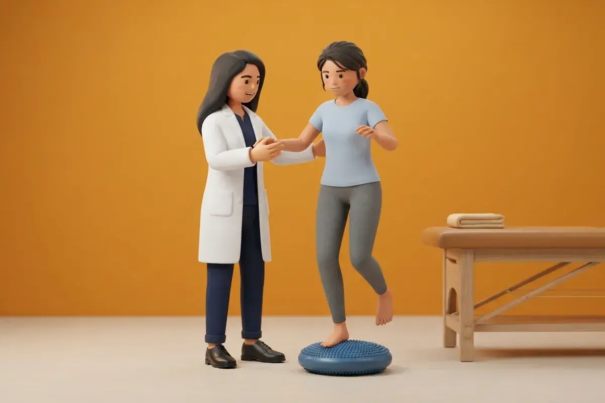
- Stage 2: Early Static and Dynamic Compensation (Days 4 to 14): Initiate active horizontal and vertical eye-head tracking drills (Cawthorne-Cooksey and VOR x1) in supported sitting, progressing to independent standing.
- Stage 3: High-Velocity Community Reintegration (Weeks 3 to 8): Progress to dynamic walking with rapid head turns, obstacle negotiation on varying surfaces, and habituation for residual visual-vestibular sensitivity.
Clinical Exercise Protocol: Early Vestibular Adaptation Drill
Early Horizontal Gaze Stabilisation (VOR x1)
Objective: Drives central neuroplastic recalibration of the vestibular nuclei to extinguish vestibular asymmetry and restore steady gaze.
- 1Sit upright in a supportive chair with armrests, holding a printed visual target 1 metre in front of your eyes.
- 2Fix your gaze securely on the target, keeping it in crisp focus.
- 3Rotate your head gently side-to-side through a 20 to 30 degree arc at a slow, comfortable speed (1 Hz).
- 4Continue for 30 to 45 seconds, resting for 1 minute if mild dizziness or nausea arises.
Clinical Comparison: Vestibular Neuritis vs Cerebellar Stroke
| Clinical Parameter | Vestibular Neuritis (Peripheral) | Cerebellar Stroke (Central) |
|---|---|---|
| Head Impulse Test | Abnormal (refixation saccade present toward lesion) | Normal (VOR pathway intact; red flag in acute vertigo) |
| Nystagmus Profile | Unidirectional horizontal-torsional | Direction-changing gaze-evoked, or pure vertical |
| Test of Skew | Absent (eyes remain aligned vertically) | Present (vertical ocular misalignment on cover test) |
| Truncal Ataxia Grade | Mild to moderate; able to sit and stand with help | Severe; completely unable to sit upright unsupported |
| Associated Neurological Signs | Absent (pure vestibular presentation) | Dysarthria, limb dysmetria, facial drop, or numbness |
Red Flags and Emergency Escalation
Under the INFARCT rule, any one of the following central signs mandates immediate emergency stroke escalation: a normal head impulse test in acute continuous vertigo, direction-changing gaze-evoked nystagmus, or vertical skew deviation on alternate cover testing. Furthermore, acute onset of severe headache, dysarthria, limb weakness, or grade 3 truncal ataxia (inability to sit upright without falling) warrants immediate tertiary hospital transfer for urgent stroke imaging.
Frequently Asked Questions
Why should dizziness tablets be stopped a few days after vestibular neuritis starts?
Medications like cinnarizine and dimenhydrinate suppress brainstem vestibular receptors. While helpful for acute vomiting, keeping them long-term stops the brain from recognizing the inner ear imbalance, preventing natural recovery.
How long does it take to fully recover from vestibular neuritis?
Severe spinning typically subsides within 3 to 5 days. With structured vestibular rehabilitation, most patients regain dynamic balance and return to full work and driving activities within 4 to 8 weeks.
Related condition guides

Related recovery guides & articles
BookPhysio.in Editorial Team
The BookPhysio.in editorial team writes and maintains this content, checking it against reliable clinical sources. Content follows our medical review policy: general information only, and no page claims a clinical review until a named registered physiotherapist has signed it off.
Read our medical review policy