
Adhesive Capsulitis (Frozen Shoulder): Freezing, Frozen and Thawing Stages and Stretching Protocols
In short
Frozen shoulder causes severe global loss of active and passive motion. Learn the 3 distinct clinical stages, capsular pattern restrictions, and stage-specific stretching protocols.
In this guide (5 sections)
Pathology of Adhesive Capsulitis
Adhesive Capsulitis, universally known as 'Frozen Shoulder', is a debilitating condition characterized by spontaneous, insidious onset of shoulder pain and progressive, painful loss of both active and passive glenohumeral range of motion.
Pathologically, it is not a simple muscle tear; it is an inflammatory fibroproliferative contracture of the glenohumeral joint capsule, characterized by high concentrations of inflammatory cytokines (TGF-beta, TNF-alpha), dense type I and III collagen deposition, and marked contracture of the rotator interval and coracohumeral ligament.
The normal axillary pouch of the capsule, which allows the arm to lift overhead, becomes densely fibrosed and obliviated, reducing intra-articular joint volume from 15 to 20 mL down to less than 5 mL.
The Three Classical Clinical Stages (Codman & Reeves)
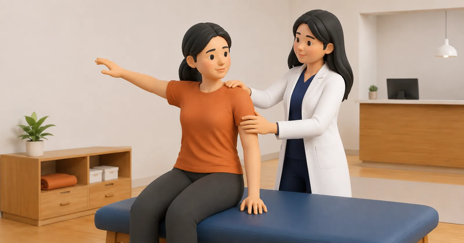
Frozen shoulder progresses through three distinct, overlapping chronological phases that require radically different physiotherapy approaches.
| Clinical Stage | Typical Duration | Clinical Presentation & Hallmark Symptoms | Physiotherapy Rehabilitation Focus | |
|---|---|---|---|---|
| 1. Freezing Phase (Painful) | 2 to 9 months | Severe, unrelenting pain; intense nocturnal waking pain; gradual loss of motion | Pain relief priority; gentle pendular swings; avoid aggressive stretching. | |
| 2. Frozen Phase (Stiff) | 4 to 12 months | Pain gradually subsides at rest; profound, rigid stiffness at end-range | Capsular stretching; end-range joint mobilizations; periscapular power. | |
| 3. Thawing Phase (Recovery) | 12 to 24+ months | Minimal pain; progressive spontaneous return of joint range of motion | Aggressive functional stretching; rotator cuff strengthening; sports return. |
The Diagnostic Capsular Pattern of the Shoulder
Adhesive Capsulitis presents with a classic Cyriax 'capsular pattern' of restriction: passive External Rotation is most severely restricted, followed by Abduction, with Internal Rotation being least restricted (ER > Abd > IR).
A critical clinical diagnostic rule: if passive external rotation with the arm at the side is normal (> 45 to 60 degrees), the diagnosis of frozen shoulder is unequivocally ruled out.
- The Golden Rule: Never Aggressively Stretch a Freezing Shoulder: During Stage 1 (freezing), aggressive forceful stretching causes reactive capsular inflammation, intensifying nocturnal pain and accelerating fibrosis. Only gentle pain-free pendular swings are permitted.
- Low-Load Prolonged Stretching (LLPS) in Stage 2: In Stage 2 (frozen), use low-load sustained stretches (cane-assisted external rotation holding at mild stretch for 60 to 90 seconds) to induce plastic deformation of collagen without tearing tissue.
- Posterior and Inferior Gliding Joint Mobilizations: Maitland Grade III and IV oscillatory joint mobilizations applied to the humeral head to stretch the posterior and inferior capsular folds.
- Sleeper Stretch and Wall Walks: Progressively walk fingers up a wall to recover overhead flexion once resting inflammation has settled.
The Diabetes Epidemic Link in India
India has one of the highest prevalences of diabetes mellitus globally. Diabetics have a 5-fold higher risk of developing frozen shoulder (up to 20 to 30 percent lifetime incidence) due to advanced glycation end-products (AGEs) cross-linking collagen fibers in the joint capsule.
In diabetic patients, frozen shoulder is frequently bilateral, more refractory to conservative care, and requires longer rehabilitation timelines.
Frequently Asked Questions
How long does a frozen shoulder take to thaw completely?
The natural history of adhesive capsulitis typically spans 18 to 24 months. Structured stage-appropriate physiotherapy significantly accelerates recovery, reducing the timeline to 6 to 12 months.
What is an ultrasound-guided hydrodilatation procedure?
Hydrodilatation (hydrodistension) involves injecting 20 to 40 mL of sterile saline and local anesthetic into the shoulder joint capsule under ultrasound guidance. The high fluid pressure gently stretches and expands the contracted capsule from the inside, providing immediate range-of-motion gains.
Why is night pain so severe in frozen shoulder?
During sleep, gravity no longer pulls the arm downward, and resting blood flow decreases, leading to venous congestion and elevated intra-capsular pressures within the inflamed, non-compliant joint capsule.
Related condition guides
Related recovery guides & articles
BookPhysio.in Editorial Team
The BookPhysio.in editorial team writes and maintains this content, checking it against reliable clinical sources. Content follows our medical review policy: general information only, and no page claims a clinical review until a named registered physiotherapist has signed it off.
Read our medical review policy