
Kyphoscoliosis-Induced Restrictive Lung Disease: Vital Capacity Loss and Pulmonary Conditioning
In short
Severe kyphoscoliosis compresses thoracic volume, producing a restrictive lung defect (FVC < 50%) that requires glossopharyngeal breathing and inspiratory muscle training.
In this guide (4 sections)
Thoracic Cavity Distortion and Restrictive Pulmonary Mechanics
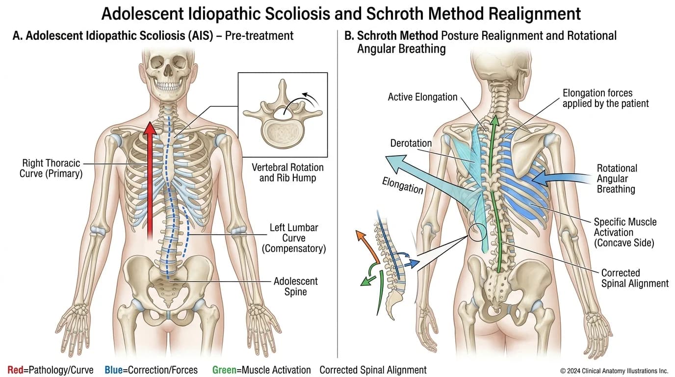
Severe thoracic spinal deformities (Cobb angle > 60 to 70 degrees, especially when combined with thoracic hyperkyphosis) cause profound distortion of the rib cage. The ribs on the convex side become crowded and rotated posteriorly, while the concave ribs collapse inward, drastically reducing total internal thoracic volume.
This anatomical distortion produces a classic Restrictive Ventilatory Defect, characterized by severe reductions in Total Lung Capacity (TLC) and Forced Vital Capacity (FVC), while the FEV1/FVC ratio remains preserved. Chest wall elastance increases up to threefold, forcing patients to adopt a rapid, shallow breathing pattern that increases dead-space ventilation and predisposes to nocturnal hypoventilation, basilar micro-atelectasis, and cor pulmonale.
Pulmonary Function Parameters and Severity Matrix in Kyphoscoliosis
| Spinal Deformity Cobb Angle | Forced Vital Capacity (FVC) | Arterial Blood Gas State | Clinical Pulmonary Risk Profile | |
|---|---|---|---|---|
| Mild Deformity (Cobb < 40 deg) | Normal or mild reduction (FVC 75-85% predicted) | Normal PaO2 and PaCO2 | Asymptomatic at rest; mild exertional breathlessness during intense sports | |
| Moderate Deformity (Cobb 40-70 deg) | Moderate restriction (FVC 60-75% predicted) | Mild exertional hypoxemia (SpO2 drops to 90%) | Impaired exercise capacity; susceptible to acute viral chest infections | |
| Severe Deformity (Cobb 70-100 deg) | Severe restriction (FVC 45-60% predicted) | Borderline awake hypercapnia; nocturnal hypoventilation | High surgical risk; requires preoperative non-invasive ventilation | |
| Very Severe (Cobb > 100 deg / Kyphosis > 90) | Extremely severe (FVC < 45% predicted) | Chronic diurnal hypercapnic respiratory failure | High risk of pulmonary hypertension and cor pulmonale (right heart failure) | |
| Inspiratory Muscle Strength | Maximal Inspiratory Pressure (MIP) < 50 cmH2O | Diaphragmatic mechanical disadvantage | Requires structured inspiratory muscle threshold loading |
Cardiopulmonary Rehabilitation and Chest Wall Expansion Protocols
- Glossopharyngeal Breathing ('Frog Breathing') Retraining: Train the patient in glossopharyngeal breathing; this technique uses the tongue and pharyngeal muscles to gulp packets of air into the lungs, stacking air above functional vital capacity to pop open collapsed micro-atelectatic lung bases.
- Inspiratory Muscle Training with Threshold Devices: Prescribe threshold inspiratory muscle training (Threshold IMT) at 30% of baseline MIP for two 15-minute sets daily; strengthening diaphragmatic fibers overcomes the high elastic resistance of the distorted rib cage.
- Concave Rib Cage Mobilization and Lateral Costal Breathing: Perform passive manual intercostal stretching and rib mobilization over the concave, sunken hemithorax during deep inspiration; physical expansion of intercostal spaces boosts vital capacity by 10% to 15%.
Frequently Asked Questions
How does severe scoliosis or a hunched back affect the lungs?
When the spine curves and twists severely (over 60 to 70 degrees), the rib cage twists along with it, shrinking the space inside your chest. Because the lungs do not have enough room to expand, they hold less air, making you feel short of breath during everyday activities.
What is 'frog breathing' (glossopharyngeal breathing) for lung expansion?
Frog breathing is a special breathing trick where you use your tongue and throat muscles to gulp small mouthfuls of air into your lungs, one after another. This gently stretches tight chest muscles and pops open collapsed air sacs that normal breaths cannot reach.
Can breathing exercises improve lung function in severe scoliosis?
Yes. While exercises cannot magically straighten a twisted rib cage, inspiratory muscle training and deep chest-expansion exercises strengthen the diaphragm and increase lung volume, significantly cutting the risk of pneumonia.
Related condition guides


Related recovery guides & articles
BookPhysio.in Editorial Team
The BookPhysio.in editorial team writes and maintains this content, checking it against reliable clinical sources. Content follows our medical review policy: general information only, and no page claims a clinical review until a named registered physiotherapist has signed it off.
Read our medical review policy