
Post-Thoracotomy and Lobectomy: Pulmonary Expansion, Chest Tube Care and Shoulder Mobility
In short
Thoracotomy and lung resection disrupt chest wall mechanics and risk frozen shoulder. Learn chest drain safety, incentive spirometry, and progressive shoulder mobilization.
In this guide (5 sections)
Surgical Impact of Thoracotomy on Pulmonary Mechanics
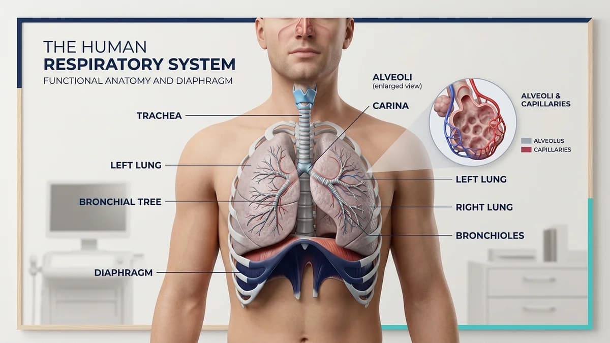
Postero-lateral thoracotomy involves dividing major chest wall muscles (latissimus dorsi, serratus anterior, intercostals) and forcefully spreading ribs to access the thoracic cavity for lung cancer resection, lobectomy, or decortication.
The procedure creates severe post-operative pain, alters chest wall kinematics, and drastically reduces Vital Capacity (VC) by up to 50 percent during the initial 48 hours.
Patients reflexively splint their hemithorax, resulting in basilar atelectasis, retention of bloody secretions, and ipsilateral shoulder protective guarding, which can rapidly progress to debilitating frozen shoulder.
Chest Drain (Intercostal Underwater Seal) Safety Guidelines
Patients emerge from thoracic surgery with one or two intercostal chest drains (ICDs) connected to an underwater seal drainage bottle to evacuate residual air, blood, and pleural fluid.
| Chest Drain Parameter | Normal Clinical Observation | Abnormal Warning Sign | Immediate Action Required | |
|---|---|---|---|---|
| 1. Fluid Oscillation (Swinging) | Fluid column rises on inspiration, falls on expiration | Absence of swinging (fluid column still) | Indicates tube blockage, kinking, or complete lung re-expansion. | |
| 2. Air Bubbling in Chamber | Intermittent bubbling during coughing or deep expiration | Continuous vigorous bubbling during tidal breathing | Indicates active bronchopleural fistula or circuit air leak; alert surgeon. | |
| 3. Drainage Color and Rate | Serosanguinous fluid decreasing to < 100 mL/day | Sudden fresh frank blood > 150-200 mL/hr | Suspect active internal thoracic hemorrhage; stop movement immediately. | |
| 4. Bottle Height Safety | Drain bottle kept strictly BELOW the level of patient chest | Lifting bottle above chest level | Accidental siphonage of pleural fluid back into chest cavity. |
Early Pulmonary Expansion and Shoulder Mobilization Protocol
Rehabilitation commences on Day 1 post-operatively with thoracic expansion exercises, wound-supported coughing, and early ipsilateral shoulder range of motion.
- Wound-Splinted Deep Breathing and SMI: The patient firmly presses a pillow over the surgical incision while taking slow deep breaths, holding for 3 seconds to re-expand remaining lung lobes.
- Ipsilateral Shoulder Active-Assisted Movements: Gentle pendular shoulder swings, cane-assisted flexion, and scapular retractions up to 90 degrees on Day 1, advancing to full overhead reaching by Day 5 to prevent capsulitis.
- Thoracic Side-Flexion and Rib Stretches: Gentle lateral trunk flexion away from the operated side to stretch tightened scar tissue and mobilize the healing intercostal space.
- Corridor Walking with Portable Drainage Carrier: Walking with the chest bottle maintained below hip level in a rolling carrier to enhance lung inflation and promote pleural air clearance.
Managing Post-Thoracotomy Pain Syndrome (PTPS)
Up to 30 percent of patients develop chronic neuropathic intercostal neuralgia (Post-Thoracotomy Pain Syndrome) from surgical rib spreader compression of the intercostal nerve.
Physiotherapists incorporate desensitization massage, Transcutaneous Electrical Nerve Stimulation (TENS) placed outside the surgical field, and myofascial release around the healing scar.
Frequently Asked Questions
Can a patient walk with a chest tube in place?
Yes. Walking with an underwater seal drain is safe and strongly encouraged, provided the bottle is kept below chest level at all times and the tubing is secured without kinking.
Why does the shoulder become stiff after lung surgery?
The latissimus dorsi and serratus anterior muscles are divided during posterolateral thoracotomy. Pain causes reflex guarding and immobilization of the shoulder girdle, leading to rapid adhesive capsulitis if active range of motion is not started on Day 1.
When can chest tubes typically be removed?
Surgeons typically remove chest drains when there is no detectable air leak for 24 hours, daily pleural fluid drainage is below 100 to 150 mL, and a chest X-ray confirms full lung expansion.
Related condition guides


Related recovery guides & articles
BookPhysio.in Editorial Team
The BookPhysio.in editorial team writes and maintains this content, checking it against reliable clinical sources. Content follows our medical review policy: general information only, and no page claims a clinical review until a named registered physiotherapist has signed it off.
Read our medical review policy