
Colles Fracture vs Smith Fracture: Dorsal vs Volar Displacement Biomechanics
In short
Differentiating dorsal Colles displacement from volar Smith displacement dictates splinting angles and exercise progression. Explore their biomechanical differences and rehabilitation principles.
In this guide (4 sections)
Distal radius fractures exhibit distinctly opposing mechanical profiles depending on wrist posture at the moment of traumatic impact. The classic Colles fracture, first described by Abraham Colles in 1814, occurs from a fall onto an outstretched hand (FOOSH) with the wrist positioned in marked dorsiflexion. This mechanism produces dorsal displacement, dorsal angulation, and radial shortening of the distal bone fragment, generating the characteristic dinner-fork deformity. Conversely, the Smith fracture, often termed the reverse Colles fracture, arises from a fall onto a flexed wrist or a direct blow to the dorsum of the hand, forcing the distal fragment into volar displacement and volar tilt. This configuration results in a garden-spade deformity with a pronounced anterior wrist prominence that creates immediate mechanical risks for the median nerve within the carpal tunnel.
Monsoon Wet Floor Falls, Two-Wheeler Skids, and Traditional Indian Cooking Rebuilding
In the Indian domestic setting, the distinction between Colles and Smith fracture mechanics corresponds to specific daily hazards. Colles fractures occur routinely when an individual slips forward on wet bathroom tiles, monsoon-soaked stone verandahs, or during sudden braking while walking on uneven footpaths, instinctively extending the palms to break the fall. Smith fractures, while less common, happen frequently during monsoon two-wheeler accidents when a scooter rider is thrown over the handlebars with hands caught in flexion, or when an individual slips backward on stairs and lands directly on the posterior aspect of the flexed wrist. Following cast removal or surgical fixation, both injuries profoundly restrict daily self-care tasks. Patients struggle to perform personal hygiene with the left hand, carry milk cans, chop firm vegetables like raw plantain, or lift traditional brass puja lamps without experiencing sharp strain at the radiocarpal junction.
Colles Malunion Mechanics, Palmer TFCC Classification, and DRUJ Instability
The biomechanical consequences of dorsal versus volar displacement differ fundamentally. In a Colles fracture, dorsal tilt creates significant tension along the volar radiocarpal ligaments and shortens the extensor tendons, leading to severe limitations in active wrist flexion and compromised finger flexion force through active insufficiency. In contrast, a Smith fracture inherently displays high mechanical instability because the dynamic pulling vector of the strong forearm flexor tendons continuously pulls the distal fragment further into the carpal tunnel. This volar displacement directly compresses the median nerve against the transverse carpal ligament, resulting in acute post-traumatic carpal tunnel syndrome in up to fifteen percent of cases. Furthermore, both patterns frequently involve avulsion of the ulnar styloid process, which compromises the dorsal or volar radioulnar ligaments of the triangular fibrocartilage complex and creates persistent rotational instability at the DRUJ.
Dart-Thrower Motion, Thermoplastic Splint Weaning, and Progressive Putty Loading
Rehabilitation must respect the original displacement vector to avoid destabilizing healing callus. For Colles fractures, initial passive and active-assisted range of motion emphasizes controlled wrist flexion and ulnar deviation, gradually restoring the lost volar angle without subjecting dorsal comminution zones to excessive tensile strain. For Smith fractures, early extension is prioritized, avoiding aggressive passive hyperflexion that could compromise healing volar cortical bone or irritate the median nerve bed. Dart-thrower motion protocols are introduced early in both conditions to re-educate the scaphoid and lunate kinematics. As bony union is confirmed radiographically, targeted soft tissue release of the pronator quadratus and flexor retinaculum is paired with progressive resistance training using graded therapeutic putty, grip dynamometers, and dumbbell curls.
Active-Assisted Wrist Extension and Flexion with Tabletop Slider
Objective: Safely restore sagittal plane wrist mobility while respecting healing bone alignment and ligament tension
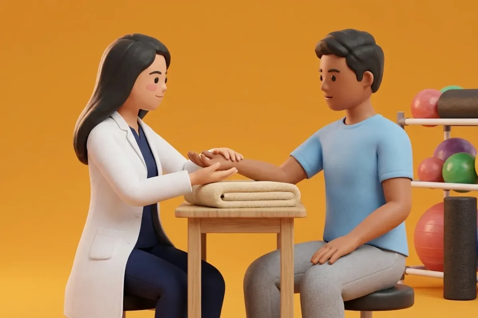
- 1Sit comfortably at a sturdy table with your affected forearm resting flat on a folded towel, wrist positioned just over the edge.
- 2Place a smooth cloth or plastic plate under your palm to eliminate frictional resistance during movement.
- 3Slowly slide your palm forward and upward into active wrist extension until you encounter a firm, comfortable stretch, holding for 5 seconds.
- 4Smoothly glide your hand backward and downward into gentle wrist flexion, using your unaffected hand to provide gentle terminal assistance without forcing through sharp pain.
- 5Return to the neutral level position, pause for 3 seconds, and ensure your fingers remain relaxed without clenching.
| Clinical Characteristic | Colles Fracture (Dorsal Displacement) | Smith Fracture (Volar Displacement) |
|---|---|---|
| Injury Mechanism | Fall onto an outstretched hand (FOOSH) with the wrist positioned in marked extension (dorsiflexion). | Fall onto a hyperflexed wrist or direct impact to the dorsal surface of the flexed hand. |
| Characteristic Deformity | Dinner-fork deformity characterized by dorsal prominence of the carpus and distal radius. | Garden-spade deformity characterized by volar prominence of the carpus and distal fragment. |
| Displacement Vector | Dorsal displacement, dorsal tilt, and radial shortening of the distal articular segment. | Volar displacement, volar tilt, and proximal displacement with frequent carpal subluxation. |
| Primary Soft Tissue Risk | Extensor pollicis longus (EPL) attrition over Lister tubercle and dorsal extensor tendon adhesions. | Acute median nerve compression within the carpal tunnel and flexor tendon impingement. |
| Rehabilitation Caution | Avoid aggressive forced passive extension in early phases; prioritize restoring lost wrist flexion. | Avoid forced end-range wrist flexion; prioritize restoring safe wrist extension and carpal clearance. |
Smith fractures carry an elevated incidence of acute median nerve palsy due to volar displacement narrowing the carpal canal. Any report of numbness, paresthesia, or severe night pain in the index and middle fingers demands immediate evaluation. For Colles fractures, evaluate the thumb tabletop lift test at every visit to rule out delayed extensor pollicis longus rupture.
Frequently Asked Questions
Why does my wrist look slightly bent even after my fracture has healed?
Residual prominence often stems from persistent dorsal or volar angulation of the bone fragment or soft tissue thickening around the distal radioulnar joint. While mild cosmetic changes are common, targeted physiotherapy focuses on restoring functional range of motion, grip strength, and pain-free forearm rotation so your daily activities remain unaffected.
Do I need a doctor referral to see a wrist fracture physiotherapist on BookPhysio.in?
BookPhysio.in does not require a physician prescription. Under the National Commission for Allied and Healthcare Professions (NCAHP) Act 2021, patients across India enjoy direct access to autonomous registered physiotherapists. Through BookPhysio.in, you connect directly with verified clinicians and pay per session with transparent fees, completely free from advance package commitments, commission markups, or platform subscription charges.
Related condition guides
Related recovery guides & articles
BookPhysio.in Editorial Team
The BookPhysio.in editorial team writes and maintains this content, checking it against reliable clinical sources. Content follows our medical review policy: general information only, and no page claims a clinical review until a named registered physiotherapist has signed it off.
Read our medical review policy