
Diaphragmatic Pacing and Phrenic Nerve Injury: Ultrasound Excursion and Ventilator Liberation
In short
Implanted diaphragmatic pacing stimulates viable phrenic motor units, enabling ventilator liberation in high cervical spinal cord injury and surgical phrenic neuropraxia.
In this guide (4 sections)
Etiology of Phrenic Nerve Injury and Diaphragmatic Dysfunction
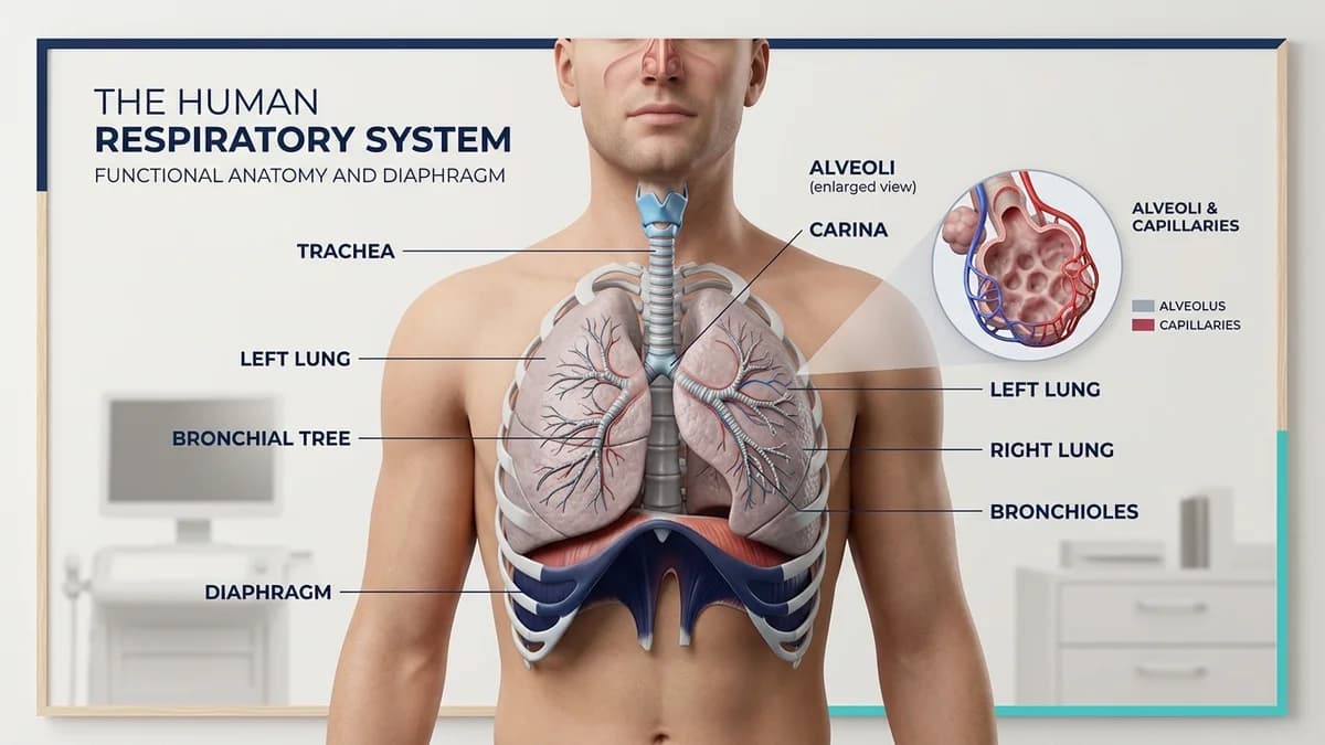
The phrenic nerve (arising from spinal roots C3, C4, and C5) provides sole motor innervation to the hemidiaphragms. Phrenic nerve injury occurs commonly following high cervical spinal cord injury (SCI), open heart surgery (due to ice slush cold injury during myocardial protection or internal mammary artery harvesting traction), or neck trauma.
Unilateral phrenic nerve palsy results in paradoxical hemidiaphragmatic elevation and a 20% to 30% loss of vital capacity, while bilateral phrenic paralysis precipitates acute, catastrophic respiratory failure and permanent ventilator dependence. Diaphragmatic Pacing Systems (DPS) offer a revolutionary neuro-prosthetic solution by electrically stimulating phrenic motor units to produce rhythmic, physiological negative-pressure breathing.
Point-of-Care Ultrasound (POCUS) and Pacing Assessment Parameters
| Diagnostic Parameter | Measurement Technique | Normal Value | Pathological / Denervation Finding | |
|---|---|---|---|---|
| Diaphragm Excursion (M-Mode) | Subcostal anterior probe angled cephalad during inspiration | 1.5 to 2.5 cm (quiet); > 5 cm (deep breath) | Zero movement or paradoxical cranial movement (< 0 cm) | |
| Diaphragm Thickness at Rest (T-di) | Zone of apposition (8th-9th ICS mid-axillary line) | 1.7 to 2.5 mm during end-expiration | < 1.5 mm (severe disuse atrophy or neurogenic wasting) | |
| Thickening Fraction (TF-di) | [(Thickness inspiration - Thickness expiration) / T-exp] x 100 | > 20% to 30% (confirms active contractility) | < 20% (predicts high risk of ventilator weaning failure) | |
| Phrenic Nerve Conduction Study | Percutaneous stimulation at posterior border of sternocleidomastoid | Latency < 9.5 ms; compound motor amplitude > 0.5 mV | Absent response indicates complete lower motor neuron transection | |
| Pacer Candidacy Requirement | Laparoscopic mapping of motor endplates | Intact lower motor neuron pathway (C3-C5) | Patients with severed phrenic motor neurons cannot be paced |
Diaphragmatic Pacing Conditioning and Weaning Protocols
- The Stepwise Pacing Endurance Progression: Begin pacemaker conditioning at 15 to 30 minutes twice daily while connected to a mechanical ventilator; atrophied type I slow-twitch diaphragmatic fibers must be conditioned slowly to avoid rapid metabolic muscle fatigue.
- Weekly Conditioning Increments: Increase pacing duration by 15 to 30 minutes per session each week as diaphragmatic thickness (measured on ultrasound) expands; advance until the patient achieves 24-hour continuous independent pacing off the ventilator.
- Postural Alignment and Abdominal Binder Support: In patients with tetraplegia, fit an elastic abdominal binder when transitioning from supine to upright wheelchair sitting; the binder replaces paralyzed abdominal wall tone, holding the flaccid diaphragm in a dome position for optimal contractility.
Frequently Asked Questions
How does a diaphragm pacemaker work to help someone breathe?
Just like a heart pacemaker regulates the heartbeat, a diaphragm pacemaker uses tiny wires attached to the breathing muscle to deliver small electrical pulses. These pulses cause the diaphragm to contract and relax smoothly, drawing air naturally into the lungs without a mechanical ventilator.
Can anyone with paralyzed breathing muscles get a diaphragm pacer?
No. The patient must have an intact phrenic nerve. If the nerve is bruised or disconnected from the brain (like in high spinal cord injury), pacing works wonders. But if the nerve itself is completely destroyed, the muscle will not respond to electrical signals.
How do doctors see if the breathing muscle is getting stronger?
Therapists and doctors use a bedside ultrasound probe on the side of your chest. They can see the diaphragm moving up and down on the screen and measure its thickness in millimeters as it rebuilds strength.
Related condition guides


Related recovery guides & articles
BookPhysio.in Editorial Team
The BookPhysio.in editorial team writes and maintains this content, checking it against reliable clinical sources. Content follows our medical review policy: general information only, and no page claims a clinical review until a named registered physiotherapist has signed it off.
Read our medical review policy