
Femoroacetabular Impingement (FAI): CAM versus Pincer Morphology, Alpha Angles, and Joint Preservation
In short
Understand the structural differences between CAM and Pincer femoroacetabular impingement, alpha angle measurement, and active physiotherapy for hip preservation.
In this guide (4 sections)
Femoroacetabular impingement, clinically designated as FAI syndrome, represents an anatomical mismatch between the proximal femoral head-neck junction and the acetabular rim, causing premature mechanical abutment during physiological hip motion. Pathomorphologically, this condition presents in two primary forms or as a combined mixed deformity. CAM morphology involves an osseous prominence or asphericity at the anterosuperior femoral head-neck junction, quantified on cross-sectional imaging or Dunn view radiographs by an elevated alpha angle exceeding 55 to 60 degrees. Conversely, Pincer morphology denotes acetabular over-coverage, resulting from general deep socket acetabular protrusion or focal cranial retroversion, frequently identified radiographically via the crossover sign. Rather than functioning as an isolated radiographic anomaly, symptomatic FAI syndrome combines structural morphology, dynamic mechanical conflict, and characteristic clinical symptoms such as deep anterior groin aching during terminal hip flexion and internal rotation. Conservative physiotherapy serves as the foundational intervention, focusing on dynamic neuromuscular joint centration and movement pattern modification to halt chondrolabral degradation.
Floor Sitting, Sukhasana Capsular Stress, and Indian Squatting Commode Realities
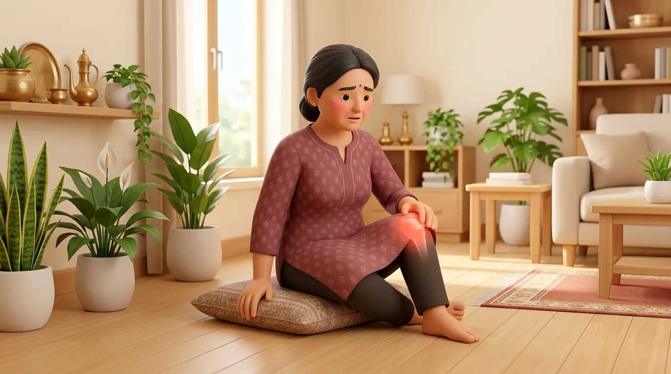
In Indian households, customary daily activities place severe mechanical demands across the hip joint capsule and labrum. Traditional postures such as sitting cross-legged on the floor (Sukhasana) or in lotus position (Padmasana) force the hip into extreme flexion, abduction, and external rotation, generating intense tensile strain on the inferior capsule while driving the femoral neck against the acetabular rim. Furthermore, using traditional Indian squatting toilets necessitates deep hip flexion exceeding 120 degrees combined with axial compression, provoking acute anterosuperior labral abutment and sharp groin pain. Preparing meals while seated on a low wooden stool (patla), washing garments in low tubs, or rising from cotton floor mattresses (gaddas) repeatedly forces the hip past its mechanical impingement threshold. Daily commuting on two-wheelers across bumpy urban roads transmits repetitive axial vibrations that aggravate reactive capsular inflammation, requiring practical ergonomic adaptations and movement pacing.
CAM and Pincer Morphology, Labral Vascularity, and Cartilage Breakdown
The progressive joint damage observed in femoroacetabular impingement stems from repetitive shear and compression stresses concentrated at the chondrolabral junction. In CAM morphology, the non-spherical femoral head slides into the acetabular socket during hip flexion, acting as an abrasive wedge that delaminates the articular cartilage from the subchondral bone, beginning at the anterosuperior acetabular margin. In contrast, Pincer morphology causes linear abutment of the normal femoral neck against the over-covering acetabular rim, crushing the fibrocartilaginous labrum and generating counter-coup chondral lesions along the posteroinferior acetabulum. The acetabular labrum features a precarious vascular architecture, wherein only the outer peripheral third receives a viable blood supply from the capsular and periosteal vessels, while the inner two-thirds constitute an avascular watershed zone. Consequently, repetitive impingement tears in this avascular region fail to heal spontaneously. Persistent mechanical shear leads to subchondral bone marrow edema, microcystic formation, and progressive articular surface degeneration that accelerates the onset of secondary hip osteoarthritis.
Isometric Abductor Strengthening, Capsular Unloading, and Low-Impact Conditioning
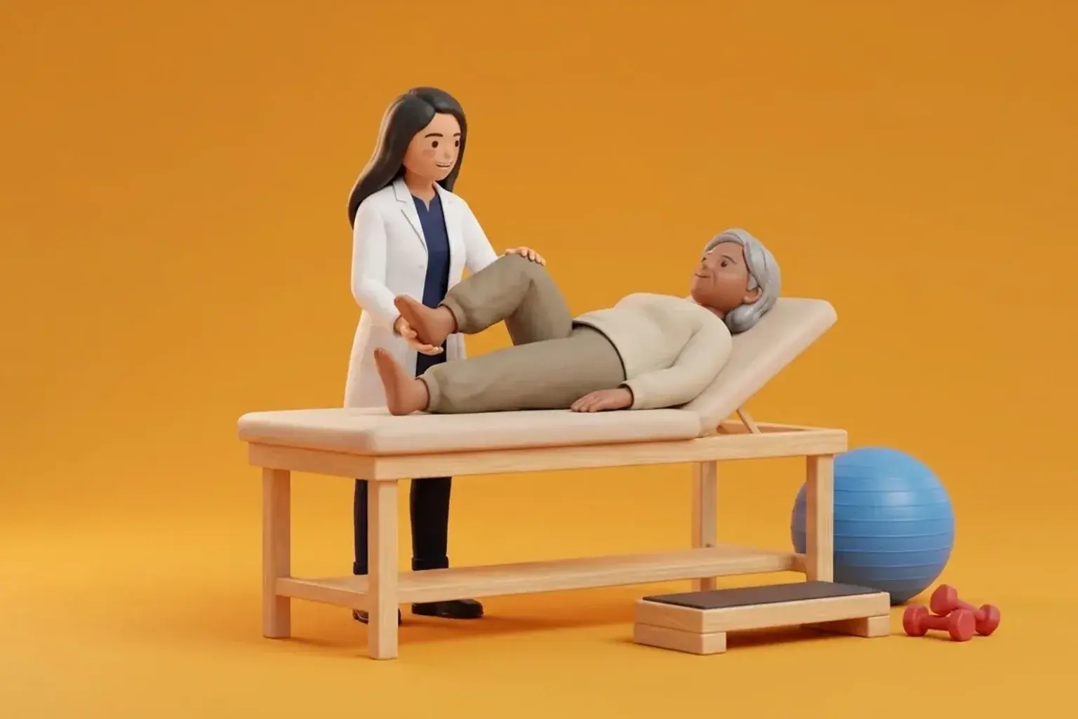
Conservative physiotherapy for femoroacetabular impingement avoids aggressive end-range stretching or forceful hip flexion mobilizations, which directly provoke bony abutment and aggravate labral distress. Modern clinical protocols prioritize restoring three-dimensional pelvic-femoral control and neuromuscular joint centration. Targeted isometric strengthening of the gluteus medius, gluteus minimus, and deep hip external rotators stabilizes the femoral head within the acetabular fossa, counteracting superior and anterior translation during dynamic movement. Pelvic tilt retraining reduces excessive anterior pelvic inclination, effectively increasing functional acetabular anteversion and clearing anterior impingement space. Aerobic conditioning transitions toward low-impact modalities such as upright stationary cycling with an elevated seat height or elliptical training, which provide rhythmic joint nutrition through synovial fluid imbibition without forcing the hip beyond 90 degrees of flexion. Manual therapy focuses on gentle inferior and lateral joint glide mobilizations to restore capsular compliance and relieve secondary muscular guarding.
Side-Lying Isometric Hip Abduction with Neutral Pelvis
Objective: Recruit the posterior fibers of the gluteus medius to improve dynamic femoral head centration without provoking anterior hip impingement
- 1Lie on your unaffected side on a firm mat, supporting your head with a pillow and aligning your spine in a straight, neutral position.
- 2Bend your bottom leg to a 45 degree angle for pelvic stability, keeping your top leg fully straight and aligned slightly behind your torso.
- 3Roll your top hip slightly forward so your pelvis does not rotate backward toward the floor.
- 4Lift your top heel 6 to 8 inches upward with your toes pointing straight forward or slightly downward, engaging your lateral gluteal muscles.
- 5Hold this elevated isometric position steadily for 10 seconds while maintaining continuous, calm abdominal breathing, then gently lower.
| Clinical Feature | CAM Morphology | Pincer Morphology |
|---|---|---|
| Primary Anatomical Deformity | Aspherical femoral head-neck junction with loss of physiological waist | Acetabular over-coverage from global depth or focal cranial retroversion |
| Radiographic Indicator | Elevated alpha angle exceeding 55 to 60 degrees on Dunn lateral radiograph | Positive crossover sign or lateral center-edge angle exceeding 40 degrees |
| Tissue Damage Mechanism | Outside-in chondral delamination followed by secondary labral detachment | Direct crushing and tearing of the labral rim with counter-coup cartilage wear |
| Rehabilitation Priority | Avoid deep flexion beyond 90 degrees and recruit posterior gluteal stabilizers | Restore neutral pelvic alignment and reduce excessive anterior pelvic tilt |
Patients presenting with persistent deep groin aching must be screened for red flag conditions. Severe, unremitting night pain that awakens you from sleep, a sudden inability to bear weight on the limb, systemic symptoms such as unexplained fevers or weight loss, or a history of high-dose oral corticosteroid therapy require urgent medical evaluation and magnetic resonance imaging to rule out femoral head avascular necrosis (AVN), occult femoral neck stress fracture, or joint space infection.
Frequently Asked Questions
Can physiotherapy resolve hip impingement without needing surgery?
Yes, structured clinical physiotherapy is the established first-line intervention for femoroacetabular impingement syndrome. While therapy does not alter underlying bone shape, it optimizes neuromuscular control, strengthens pelvic stabilizers, and teaches movement modifications that eliminate mechanical conflict between the femur and acetabular rim. Clinical studies demonstrate that a majority of patients achieve significant pain relief, improved mobility, and enhanced functional independence through targeted conservative rehabilitation.
Do I need a doctor referral to see a hip physiotherapist on BookPhysio.in?
BookPhysio.in does not require a doctor referral. Under the National Commission for Allied and Healthcare Professions (NCAHP) Act 2021, patients across India enjoy direct access to autonomous registered physiotherapists. Through BookPhysio.in, you connect directly with verified clinicians and pay per session with transparent fees, completely free from advance package commitments, commission markups, or platform subscription charges.
Related condition guides
Related recovery guides & articles
BookPhysio.in Editorial Team
The BookPhysio.in editorial team writes and maintains this content, checking it against reliable clinical sources. Content follows our medical review policy: general information only, and no page claims a clinical review until a named registered physiotherapist has signed it off.
Read our medical review policy