
Heel Lift Inserts and Drop Height Elevation in Achilles Tendinopathy Rehabilitation
In short
Learn how heel lift inserts and footwear drop height elevation reduce Achilles tendon tensile and compressive strain, accelerating early rehabilitation.

In this guide (4 sections)
In the early management of both midportion and insertional Achilles tendinopathy, modifying mechanical tissue stress is essential to settling acute symptoms and creating a favorable environment for active rehabilitation. Among simple conservative interventions, temporary heel lift inserts and footwear drop height adjustments provide immediate, clinically verified mechanical unloading. Biomechanically, introducing a raised heel elevates the calcaneus relative to the forefoot, effectively placing the talocrural joint in slight plantarflexion. This elevation shortens the functional distance between the origin of the gastrocnemius-soleus complex and the calcaneal insertion, markedly reducing passive tensile strain across the tendon during the midstance and terminal stance phases of gait. For insertional pathology, heel lifts serve an even more critical role by physically preventing the anterior tendon fibers from compressing against the posterosuperior calcaneal ridge.
Transitioning from Traditional Flat Footwear to Supportive Elevated Profiles
The prevalence of zero-drop and negative-heel footwear in Indian daily life explains why Achilles tendinopathy frequently flares and proves resistant to healing. Countless individuals walk barefoot on residential tiles, wear ultra-flat leather Kolhapuris, or use wafer-thin rubber slippers that provide zero millimetres of heel elevation. When someone with an inflamed Achilles tendon walks completely flat, the ankle must dorsiflex ten to fifteen degrees with every step, subjecting the tendon to maximal tensile stretch and severe insertional compression. Conversely, many Indian women who habitually wear heeled sandals or ethnic wedges for years experience severe Achilles pain when abruptly switching to zero-drop walking shoes or flat walking slippers for morning walks. Introducing bilateral, firm silicone or high-density cork heel lifts (8 to 12 millimetres) into daily shoes immediately cushions the heel and creates a safe mechanical buffer, allowing daily domestic tasks to be performed without triggering acute tendon flares.
Achilles Tensile Strain Reduction, Moment Arm Mechanics, and Offloading Kinetics
The mechanical benefits of heel lifts have been quantified extensively through kinematic motion analysis and in-vivo tendon force transducers. Studies confirm that a twelve-millimetre heel lift reduces peak Achilles tendon tensile strain by twelve to fifteen percent during normal walking gait. Furthermore, elevating the heel shifts the ground reaction force vector slightly forward relative to the ankle joint center of rotation, shortening the external dorsiflexion moment arm and reducing the internal muscular force demand on the triceps surae to stabilize the tibia. In insertional tendinopathy and retrocalcaneal bursitis, this slight plantarflexion angle prevents the tendon from entering the critical impingement zone where soft tissue is crushed against the calcaneal tuberosity. Importantly, heel lifts must always be inserted bilaterally, even if symptoms are unilateral, to prevent pelvic obliquity and compensatory gait deviations that could provoke secondary knee or lower back symptoms.
Material Selection, Bilateral Placement, and Graduated Weaning Timelines
For maximum therapeutic efficacy, heel lift selection and implementation must follow strict biomechanical parameters. Soft, squishy foam or gel pads are strongly discouraged because they compress unpredictably under bodyweight, creating mechanical instability that forces the calf to over-contract to maintain balance. Instead, clinicians prescribe firm, high-density viscoelastic silicone, ethylene-vinyl acetate (EVA), or solid cork lifts measuring between 8 and 12 millimetres in height. The inserts must be worn in all closed footwear throughout the day, including supportive indoor house shoes. However, heel lifts represent a temporary rehabilitation crutch, not a permanent orthotic dependency. As progressive strengthening restores tendon load tolerance over eight to twelve weeks, the heel lifts are systematically weaned down in two to three-millimetre decrements every two weeks until normal plantigrade mechanics are fully restored without symptom recurrence, ensuring durable long-term functional recovery and robust tissue tolerance during barefoot standing.
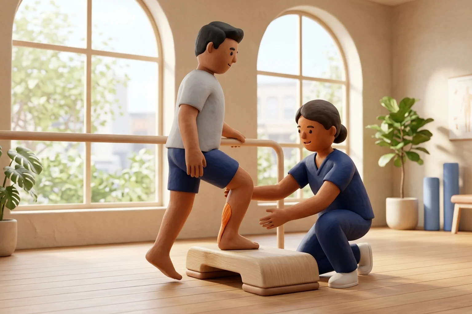
Bilateral Controlled Heel-Lift Supported Balance and Weight Shift
Objective: Condition proprioceptive stability and gradual loading with mechanical heel-lift assistance
- 1Place a matched pair of 10 mm firm heel lifts securely inside a supportive pair of lace-up running shoes.
- 2Lace your shoes securely and stand tall with feet hip-width apart on a level, stable floor.
- 3Gently shift seventy percent of your bodyweight onto the affected leg, maintaining an upright pelvis and soft knee alignment.
- 4Hold this stable single-leg weight-bearing posture for twenty seconds, feeling the calf muscles engage without sharp posterior heel tension.
- 5Shift your weight smoothly back to the center, rest for ten seconds, and alternate sides for the prescribed volume.
| Biomechanical Metric | Zero-Drop / Flat Footwear | 8 to 12 mm Firm Heel Lift | Clinical Relevance in Rehabilitation |
|---|---|---|---|
| Peak Achilles Tensile Strain | Maximal tensile strain during midstance and push-off | 12% to 15% reduction in peak tensile strain | Immediate relief of reactive midportion tensile overload |
| Entheseal Impingement Risk | High compressive crush against calcaneal bone | Substantial reduction in bone-on-tendon compression | Mandatory protection for insertional tendinopathy and bursitis |
| Calf Muscular Workload | Elevated internal stabilizing torque required | Slightly reduced muscular demand, protecting fatigue | Enables patient to perform daily walking without flares |
| Gait Symmetry Requirement | Symmetrical flat contact across both lower limbs | Must be worn bilaterally to prevent pelvic tilt | Prevents secondary hip, knee, or sacroiliac pain |
While heel lifts are invaluable during the reactive phase of Achilles tendinopathy, leaving them in place indefinitely without a planned weaning schedule can cause chronic adaptive shortening of the gastrocnemius-soleus complex and permanent loss of talocrural dorsiflexion mobility. Once a patient achieves four consecutive weeks of pain-free walking and demonstrates twenty unilateral calf raises, the heel lift height must be systematically reduced by two to three millimetres every fourteen days under physiotherapy guidance until completely removed.
Frequently Asked Questions
Can I wear a heel lift in only one shoe if only one of my heels hurts?
No, you must never wear a heel lift in only one shoe. Inserting an 8 to 12-millimetre lift into only the symptomatic shoe creates an artificial leg-length discrepancy. This tilt alters your pelvic alignment, increases shear stress across your sacroiliac joints, and causes compensatory twisting at the knees and lumbar spine. Always place identical, matched heel lifts in both shoes simultaneously to ensure your pelvis and spine remain completely level and balanced during walking.
Do I need a doctor referral to treat Achilles tendinopathy on BookPhysio.in?
BookPhysio.in does not require a doctor referral. Under the National Commission for Allied and Healthcare Professions (NCAHP) Act 2021, patients across India enjoy direct access to autonomous registered physiotherapists. Through BookPhysio.in, you connect directly with verified clinicians and pay per session with transparent fees, completely free from advance package commitments, commission markups, or platform subscription charges.
Related condition guides
Related recovery guides & articles
BookPhysio.in Editorial Team
The BookPhysio.in editorial team writes and maintains this content, checking it against reliable clinical sources. Content follows our medical review policy: general information only, and no page claims a clinical review until a named registered physiotherapist has signed it off.
Read our medical review policy