
Malignant Pleural Effusion: Indwelling Pleural Catheters (PleurX), Drainage, and Mobility Safety
In short
Patients with indwelling tunneled pleural catheters (PleurX) can walk and mobilize safely by adhering to catheter anchoring, drainage timing, and avoiding sudden traction.

In this guide (4 sections)
Tumor Lymphatic Blockade, Pleural Permeability, and MPE Pathophysiology
Malignant Pleural Effusion (MPE) is a frequent and distressing manifestation of advanced metastatic cancer, most commonly originating from lung cancer, breast carcinoma, ovarian adenocarcinoma, and malignant pleural mesothelioma. In healthy individuals, approximately 15 to 20 mL of serous fluid lubricates the pleural space, maintained by a balance between parietal pleural capillary filtration and lymphatic drainage. Metastatic tumor implants on the pleural surfaces disrupt this homeostasis through two synergistic mechanisms.
Tumor cells release high levels of Vascular Endothelial Growth Factor (VEGF), dramatically increasing pleural capillary microvascular permeability and protein exudation. Concurrently, tumor emboli obstruct the subpleural lymphatic stomata of the parietal pleura, abolishing normal lymphatic clearance. Massive exudative fluid accumulations (often 1 to 2 liters) compress the adjacent lung parenchyma (compressive atelectasis), displace the mediastinum, flatten the diaphragm, and induce severe, progressive dyspnea, dry cough, and chest wall heaviness.
Standardized Clinical Outcome Measures and Catheter Management
| Clinical Assessment Domain | Standard Monitoring Method | Expected Clinical Finding | Rehabilitation & Safety Mandate | |
|---|---|---|---|---|
| Vacuum Bottle Drainage Volume | Intermittent vacuum bottle connection (typically 500 to 1,000 mL) | Spontaneous fluid drainage relieves lung compression | DRAIN FLUID 30-60 MINUTES PRIOR TO EXERCISE; maximizes lung expansion | |
| Re-Expansion Pulmonary Pain | Pain scale during final phase of vacuum bottle drainage | Sharp, aching chest pain secondary to visceral-parietal pleural touch | Pause vacuum drainage if pain exceeds 4/10 or coughing occurs | |
| Catheter Exit Site & Cuff Integrity | Visual inspection of subcutaneous polyester Dacron cuff | Cuff well-anchored under skin; exit site clean, dry, zero redness | Ensure catheter is coiled and taped flat; NO TRACTION ON TUBE | |
| Exertional Pulse Oximetry (SpO2) | Continuous SpO2 measurement during hallway ambulation | Improvement in SpO2 from < 90% pre-drainage to > 94% post-drainage | Prescribe progressive corridor walking post-drainage | |
| Pleural Infection / Empyema Screen | Inspection of drained fluid clarity and patient temperature | Fluid should be straw-colored or serosanguinous; afebrile | Cloudy, purulent fluid with fever indicates empyema; STAT hospital referral |
Standardized Clinical Rehabilitation Protocols and Movement Rules
- Optimal Timing: Mobilize Immediately Post-Pleural Drainage: Schedule physiotherapy and walking drills 30 to 60 minutes following vacuum bottle fluid drainage; evacuating 500 to 1,000 mL of pleural fluid immediately decompresses collapsed lung alveoli and unweights the diaphragm, giving the patient their peak respiratory window for exercise.
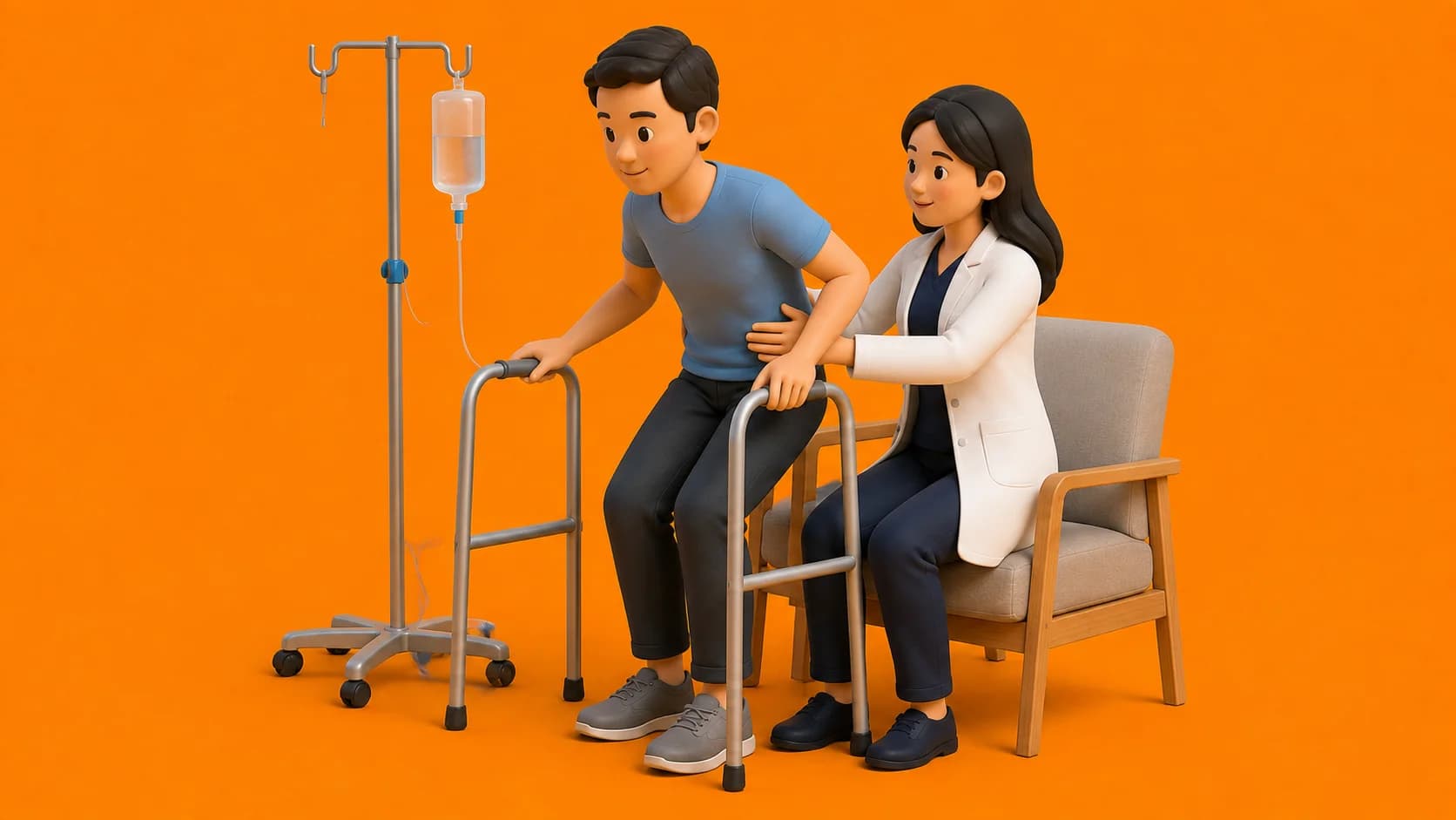
- Meticulous Catheter Coiling and Tension-Free Taping: Inspect the PleurX catheter before any transfers; ensure the external silicone tube is gently looped without acute kinks and taped securely beneath a transparent waterproof dressing; never allow clothing or gait belts to pull on the catheter exit site.
- Active Thoracic Expansion and Deep Diaphragmatic Retraining: Teach lateral costal expansion and gentle sustained maximal inspirations; expanding the underlying lung encourages the visceral and parietal pleura to come into apposition, facilitating spontaneous pleurodesis (natural fusion of the pleural layers) over time.
- Strict Water Submersion Prohibition: Strictly prohibit swimming pools, bathtubs, and hot tubs; having a tunneled catheter entering the pleural cavity creates an open conduit for aquatic bacteria to cause catastrophic pleural empyema.
Frequently Asked Questions
What is a PleurX catheter and how does it help with cancer fluid around the lung?
A PleurX catheter is a small, soft silicone tube placed between your ribs into the chest space with the end resting under your skin. Instead of going to the hospital for painful needle taps every week, you or a nurse can connect a small vacuum bottle at home to gently drain fluid whenever you feel breathless.
Can I walk and exercise with a drainage tube in my chest?
Yes, absolutely! The tube is taped securely flat against your chest under a soft bandage. In fact, doing your walking exercises right after draining fluid is ideal because your lung has just expanded and you can breathe with much greater ease.
Why does my chest hurt toward the end of draining the fluid bottle?
As the bottle finishes pulling the fluid out, your lung expands all the way and its outer surface gently touches the inside of your ribs. This contact often causes a sharp, temporary ache or makes you cough. Simply clamp the tube when the ache begins; it will settle within minutes.
Related condition guides

Related recovery guides & articles
BookPhysio.in Editorial Team
The BookPhysio.in editorial team writes and maintains this content, checking it against reliable clinical sources. Content follows our medical review policy: general information only, and no page claims a clinical review until a named registered physiotherapist has signed it off.
Read our medical review policy