
Midportion versus Insertional Achilles Tendinopathy: Differential Pathology and Biomechanics
In short
Examine the critical anatomical and mechanical distinctions between midportion and insertional Achilles tendinopathy, from vascular watershed zones to entheseal compressive shear.

In this guide (4 sections)
Achilles tendinopathy presents along two distinct anatomical, biomechanical, and histopathological entities that require fundamentally divergent clinical management: midportion tendinopathy and insertional tendinopathy. The Achilles tendon is the thickest and strongest tendon in the human body, transmitting tensile loads up to twelve times body weight during sprinting and jumping. Midportion tendinopathy develops two to six centimetres proximal to the calcaneal insertion, an anatomical region known as the relative watershed zone due to its limited microvascular perfusional supply from the posterior tibial artery branches. In contrast, insertional tendinopathy occurs directly at the enthesis within two centimetres of the calcaneal tuberosity, where the tendon blends with the periosteum, adjacent retrocalcaneal bursa, and fibrocartilage. Conflating these two presentations frequently leads to clinical failure, because standard eccentric exercises that successfully rehabilitate midportion lesions often aggravate insertional pathology through excessive compressive impingement.
Barefoot Marble Floors, Flat Kolhapuris, and Indian Household Morning Stiffness
In Indian households, domestic footwear habits and architectural features dramatically amplify the mechanical stressors governing Achilles tendinopathy. Walking barefoot across unyielding marble, granite, or vitrified tile flooring forces the foot into maximal plantigrade contact, depriving the heel of cushioning and increasing dorsiflexion demands during late midstance. Many individuals habitually wear traditional Kolhapuri chappals, thin-soled flat leather sandals, or worn rubber slippers (hawai chappals) that possess negative heel-to-toe drop heights and rigid footbeds. When an inflamed tendon is subjected to these zero-drop surfaces, the tensile strain rises precipitously. Patients consistently describe debilitating morning first-step pain upon getting out of bed, where taking the first ten paces across cold bedroom tiles provokes sharp lancinating pain behind the heel. Furthermore, operating heavy manual gear two-wheelers in congested metropolitan traffic requires forceful kick-starting and repeated sudden foot-down stops, introducing abrupt eccentric shock to unprepared Achilles fibers. This repeated shock rapidly triggers inflammatory flares in patients attempting to commute to work.
Tensile Overload versus Entheseal Compressive Shear and Fibrocartilaginous Metaplasia
The histopathology of midportion Achilles tendinopathy is predominantly driven by excessive tensile loading that exceeds the physiological adaptive threshold of tenocytes. This triggers a continuum beginning with reactive tendinopathy, progressing to tendon dysrepair, and culminating in degenerative tendinopathy characterized by hypercellularity, collagen type I disorganization, abundant glycosaminoglycan accumulation, and neovascular capillary ingrowth accompanied by sensory nerve sprout proliferation. Conversely, insertional tendinopathy is characterized by combined tensile and compressive shear forces. As the ankle moves into dorsiflexion past neutral, the deep anterior aspect of the Achilles tendon is compressed directly against the superior tuberosity of the calcaneus and the retrocalcaneal bursa. Under repetitive compressive stress, tenocytes undergo chondroid metaplasia, producing aggrecan and transforming normal tensile tendon matrix into vulnerable fibrocartilaginous tissue that frequently develops calcific enthesophytes and bone spurs. This mechanical conflict impairs blood supply and triggers persistent pain during weight-bearing activities, requiring specific interventions that eliminate bone-on-tendon compressive contact.
Zonal Loading Protocols, Flat-Floor Adaptation, and Kinetic Chain Strengthening
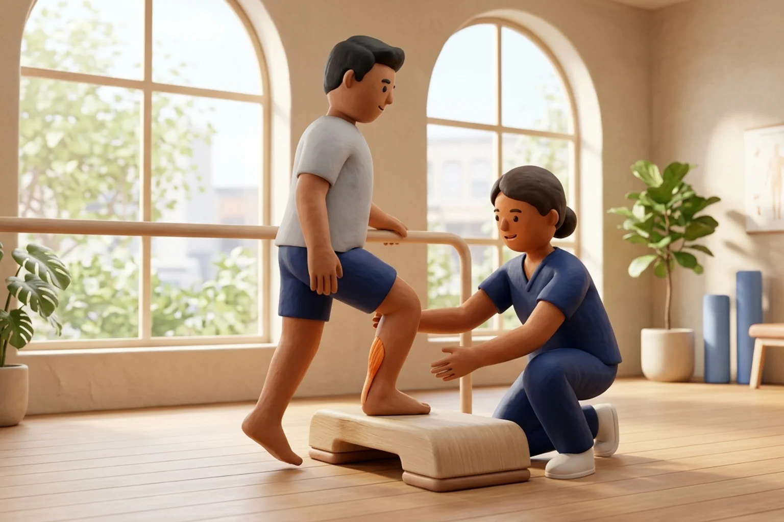
Rehabilitation must be rigorously customized to the anatomical zone of involvement. For midportion tendinopathy, the classic Alfredson protocol incorporates full-range eccentric heel drops moving from full plantigrade elevation down into deep dorsiflexion over the edge of a step. This tensile elongation under load stimulates collagen realignment and reverses neovascular proliferation. However, performing eccentric heel drops off a step in insertional tendinopathy is strongly contraindicated because drop-depth dorsiflexion directly impinges the enthesis against the calcaneus. Instead, insertional rehabilitation must be executed strictly on flat ground, maintaining the ankle between neutral dorsiflexion and terminal plantarflexion. Both conditions necessitate progressive heavy slow resistance training to rebuild triceps surae force capacity, alongside targeted soleus isolation, hip abductor conditioning, and graduated cadence modification. Restoring symmetrical calf power and ankle mobility ensures sustainable return to daily activities and active recreational pursuits, protecting the remodeled tendon matrix from premature re-injury during high-velocity locomotive tasks.
Flat-Ground Isometric Calf Raise Hold with Neutral Ankle Alignment
Objective: Induce tendon neuroplastic analgesia and build triceps surae tension without entheseal compressive impingement
- 1Position yourself standing barefoot or in supportive flat trainers on a completely level floor, lightly resting your fingertips against a sturdy wall or countertop for balance.
- 2Distribute your body weight evenly across the balls of both feet, keeping your knees fully extended and your spine upright in neutral posture.
- 3Press firmly through the first and second metatarsal heads to elevate your heels approximately two to three inches off the floor into a mid-range calf raise.
- 4Hold this elevated plantarflexed position statically for thirty to forty-five seconds, ensuring your ankles do not roll outward into supination.
- 5Slowly lower your heels back to the flat floor over a controlled three-second count, rest for forty-five seconds, and repeat for the prescribed volume.
| Pathological Feature | Midportion Tendinopathy | Insertional Tendinopathy | Physiotherapy Implication |
|---|---|---|---|
| Anatomical Location | 2 to 6 cm proximal to calcaneus in watershed zone | Distal 2 cm at calcaneal insertion and enthesis | Guides palpation site and imaging ultrasound focus |
| Primary Mechanical Driver | Pure high-tensile energy storage and release cycles | Combined tensile load and dorsiflexion compression | Determines whether step drop-depth is allowed |
| Retrocalcaneal Bursa | Rarely involved or entirely normal | Frequently inflamed with retrocalcaneal bursitis | Requires anti-compressive footwear modifications |
| Eccentric Drop Depth | Permitted into deep dorsiflexion over step edge | Strictly restricted to flat floor to avoid impingement | Essential safety boundary to prevent symptom aggravation |
Before commencing any eccentric or heavy loading protocol, clinicians must systematically screen for recent oral fluoroquinolone antibiotic consumption (such as ciprofloxacin, levofloxacin, or moxifloxacin) within the preceding twelve months, as well as local corticosteroid injections. Fluoroquinolones induce direct tenocyte toxicity, matrix metalloproteinase upregulation, and extensive collagen degradation, increasing the hazard of sudden macroscopic tendon rupture. If a history of fluoroquinolone exposure is established, aggressive high-load eccentric drills must be suspended in favor of gentle low-load isometrics, accompanied by prompt orthopaedic consultation.
Frequently Asked Questions
Why does my heel hurt worse when I perform traditional calf stretches against a wall?
Traditional runner wall stretches force the ankle into extreme passive dorsiflexion. In insertional Achilles tendinopathy, this extreme dorsiflexion physically crushes the deep anterior fibers of the tendon against the rigid posterosuperior corner of the heel bone (calcaneus). Instead of loosening the tendon, you are generating severe compressive shear stress across an already sensitized enthesis. Discontinue all dropped-heel and wall stretches immediately, and transition to flat-surface isometric calf exercises under the guidance of a qualified physiotherapist.
Do I need a doctor referral to treat Achilles tendinopathy on BookPhysio.in?
BookPhysio.in does not require a doctor referral. Under the National Commission for Allied and Healthcare Professions (NCAHP) Act 2021, patients across India enjoy direct access to autonomous registered physiotherapists. Through BookPhysio.in, you connect directly with verified clinicians and pay per session with transparent fees, completely free from advance package commitments, commission markups, or platform subscription charges.
Related condition guides
Related recovery guides & articles
BookPhysio.in Editorial Team
The BookPhysio.in editorial team writes and maintains this content, checking it against reliable clinical sources. Content follows our medical review policy: general information only, and no page claims a clinical review until a named registered physiotherapist has signed it off.
Read our medical review policy