
Anterior (Superior) Canal BPPV: Downbeating Torsional Nystagmus & Deep Head-Hanging (Yacovino) Maneuver
In short
Anterior canal BPPV is rare and notoriously tricky, producing downward-beating nystagmus that mimics central brainstem lesions. Learn the Yacovino repositioning procedure.
In this guide (5 sections)
Rarity and Anatomy of Anterior Semicircular Canal BPPV
Anterior (superior) semicircular canal BPPV (AC-BPPV) represents approximately 1 to 3 percent of all diagnosed BPPV cases. Because the anterior canal's highest anatomical point (the apex) is oriented superiorly, free-floating otoliths naturally tend to fall downward out of the canal back into the common crus or utricle under resting gravity.
However, following severe head trauma, whiplash, or vigorous inversion exercises (such as headstands or downward dog in yoga), otoconia can become trapped in the anterior canal, triggering vertigo upon looking down or bending forward.
Diagnostic Presentation: Downbeating Torsional Nystagmus
When performing a Dix-Hallpike test or straight head-hanging test, anterior canal BPPV produces a paroxysmal downbeating nystagmus, usually accompanied by a subtle torsional component beating toward the affected ear.
Because downbeating nystagmus is also a hallmark of dangerous central neurological pathologies (such as cerebellar stroke, Chiari malformation, or multiple sclerosis), the clinician must meticulously differentiate benign peripheral BPPV from central vestibular emergencies.
| Clinical Parameter | Peripheral Anterior Canal BPPV | Central Neurological Lesion (Cerebellar/Brainstem) | |
|---|---|---|---|
| Onset Latency | Definite latency (2 to 8 seconds delay) | Immediate onset with zero latency | |
| Duration | Transient paroxysmal burst (< 45 to 60 seconds) | Persistent; continuous as long as position held | |
| Fatiguability | Nystagmus fatigues on repeated testing | Zero fatigue on repeated testing | |
| Fixation Suppression | Suppressed by visual fixation in well-lit room | Not suppressed by visual fixation; enhanced | |
| Associated Signs | Isolated rotational vertigo and nausea | Dysmetria, ataxia, dysarthria, cranial nerve palsies |
The Deep Head-Hanging (Yacovino) Maneuver
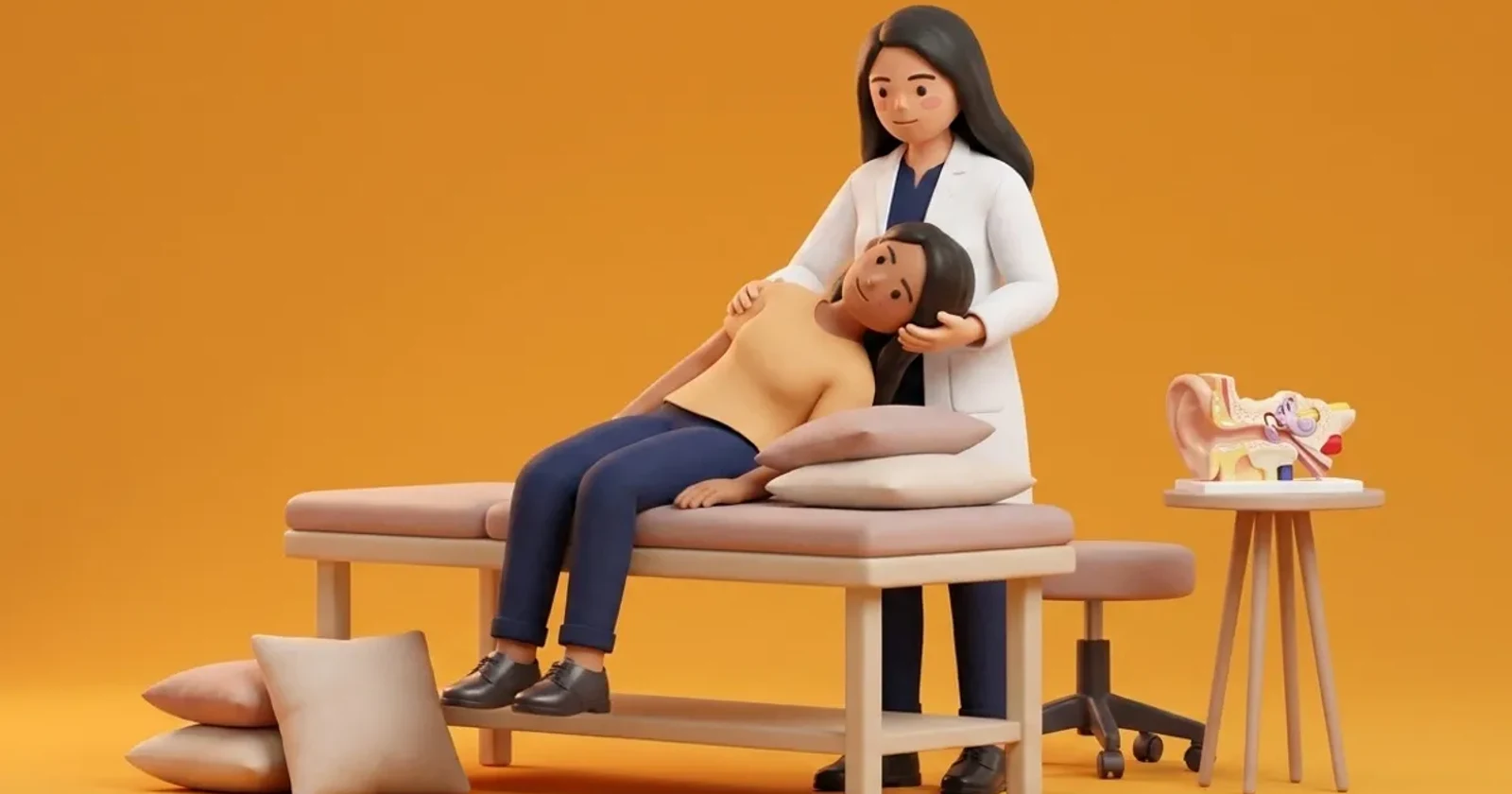
Developed by Dr. Aquiles Yacovino, this maneuver has the distinct advantage of successfully clearing anterior canal debris from either the left or right ear without requiring definitive identification of which side is affected.
The maneuver relies on symmetric sagittal plane head hanging and rapid flexion.
- Step 1: Straight Deep Head-Hanging: Lower patient straight back into supine with head hanging backward at least 30 degrees below horizontal; hold for 30 seconds.
- Step 2: Rapid Cervical Chin Tuck: Rapidly flex the neck forward, bringing the chin firmly against the sternum while remaining supine; hold for 30 seconds.
- Step 3: Return to Upright Sitting: Help the patient sit smoothly upright while maintaining chin-to-chest flexion for 30 seconds before lifting head to neutral.
The Reverse Epley Alternative
When the affected ear is definitively identified via video-oculography, a 'Reverse Epley' maneuver can be performed, mirroring the standard Epley sequence in reverse to navigate the anterior canal arch.
Frequently Asked Questions
Why is anterior canal BPPV so easily mistaken for a brain problem?
Downbeating nystagmus is typically caused by cerebellar or brainstem lesions. Clinicians must observe the classic BPPV latency, crescendo-decrescendo curve, and fatigue to safely confirm peripheral canalithiasis.
How does the Yacovino maneuver work if we do not know which ear is affected?
Because the anterior canals are oriented in symmetrical sagittal planes that meet at the common crus, straight backward head hanging and chin tucking moves debris through either anterior canal back into the utricle simultaneously.
Can inverted yoga postures cause anterior canal BPPV?
Yes. Sustained inversions like headstands (Sirsasana) place the anterior semicircular canals in an inverted, gravity-dependent position where loose utricular debris can tumble into the canal.
Related condition guides


Related recovery guides & articles
BookPhysio.in Editorial Team
The BookPhysio.in editorial team writes and maintains this content, checking it against reliable clinical sources. Content follows our medical review policy: general information only, and no page claims a clinical review until a named registered physiotherapist has signed it off.
Read our medical review policy