
Proximal Humerus Fracture PHILOS Plate ORIF: Neer Classification and Codman Pendular Rehab
In short
Proximal humerus fractures in osteoporotic older adults require anatomical locking fixation. Learn the Neer classification, PHILOS plate biomechanics, and phased shoulder rehab.
In this guide (5 sections)
The Anatomy of Proximal Humerus Fractures
Proximal humerus fractures represent the third most common non-vertebral osteoporotic fracture in older adults (after hip and distal radius fractures), predominantly affecting postmenopausal women following a low-energy fall from standing height.
Anatomically, fractures divide the proximal humerus into four classic segments (Neer's classification): the humeral head articular segment, the greater tuberosity (rotator cuff attachment), the lesser tuberosity (subscapularis attachment), and the humeral shaft.
Unstable, displaced 2-part, 3-part, or 4-part fractures undergo Open Reduction and Internal Fixation (ORIF) with a Proximal Humeral Internal Locking System (PHILOS) plate, which provides angular stable fixation in osteoporotic bone.
The Neer Classification and Tuberosity Union
Neer defined a fracture 'part' as displaced only if there is > 1.0 cm of displacement or > 45 degrees of angulation.
| Neer Fracture Category | Number of Displaced Segments | Vascular Head Risk (AVN) | Recommended Treatment Strategy | |
|---|---|---|---|---|
| 1. One-Part (Non-displaced) | Any number of fracture lines, but < 1 cm displacement | Very low AVN risk (< 3%) | Conservative: sling for 2-3 weeks; early Codman pendular swings. | |
| 2. Two-Part Fracture | One segment displaced (e.g., surgical neck or greater tuberosity) | Low to moderate AVN risk | PHILOS plate ORIF or closed reduction and percutaneous pinning. | |
| 3. Three-Part Fracture | Two segments displaced (e.g., shaft + greater tuberosity) | Moderate AVN risk (15 to 25%) | PHILOS locking plate with rotator cuff suture cerclage. | |
| 4. Four-Part Fracture | All four segments displaced; humeral head dislocated | High AVN risk (> 50%) in elderly | Reverse Total Shoulder Arthroplasty (rTSA) in osteoporotic elderly. |
Phase-Wise Post-Operative Rehabilitation
The primary clinical objective is balancing early passive mobilization to prevent frozen shoulder with protecting the healing tuberosities and bone plate construct.
- Phase 1: Suture & Sling Protection (Weeks 0 to 4): Continuous sling wear; perform active finger, wrist, and elbow motion on Day 1; begin gentle Codman pendular swings leaning forward; passive forward flexion up to 90 degrees in scapular plane.
- Phase 2: Active-Assisted Elevation (Weeks 5 to 8): Wean from sling; begin active-assisted movements (supine cane exercises, overhead pulleys) advancing forward flexion to 120 degrees; external rotation strictly to 30 degrees to protect the repaired tuberosity.
- Phase 3: Active Range of Motion & Isometrics (Weeks 9 to 12): Full active unassisted elevation against gravity; begin submaximal isometric rotator cuff strengthening once radiographic callus bridging is confirmed.
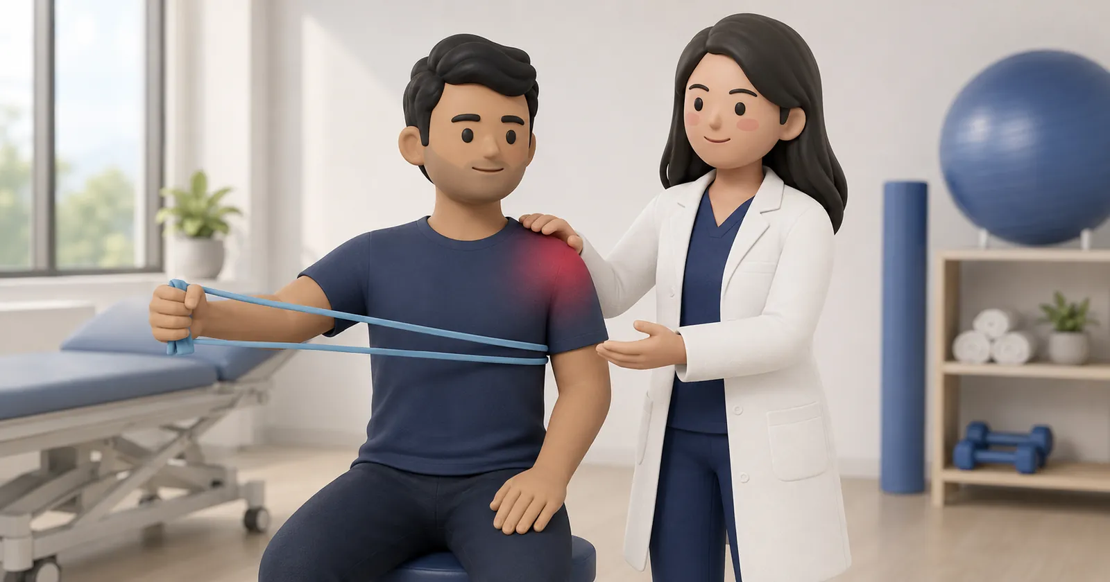
- Phase 4: Functional Resistance Training (Months 3 to 6): Progressive resistance band training, light dumbbell exercises, and functional household reaching.
Avascular Necrosis (AVN) Monitoring
Disruption of the ascending branch of the anterior humeral circumflex artery can lead to avascular necrosis of the humeral head, typically manifesting 6 to 18 months post-injury with collapse and deep joint aching.
Physiotherapists monitor follow-up radiographs and adjust loading accordingly.
Frequently Asked Questions
What are Codman's pendular exercises?
The patient bends forward 90 degrees at the waist, supporting themselves on a table with the good arm, allowing the injured arm to hang down like a pendulum. By gently swaying the trunk in circles, the arm moves passively in the socket with zero active shoulder muscle contraction.
How long does a proximal humerus fracture take to heal?
Bony union typically occurs within 6 to 8 weeks, with complete remodeling and solid trabecular consolidation taking 3 to 6 months.
Why is external rotation restricted after greater tuberosity fracture fixation?
The infraspinatus and teres minor tendons attach to the greater tuberosity. Aggressive external rotation stretches these tendons, creating high tensile forces that can pull the healing bone fragment off the humerus.
Related condition guides


Related recovery guides & articles
BookPhysio.in Editorial Team
The BookPhysio.in editorial team writes and maintains this content, checking it against reliable clinical sources. Content follows our medical review policy: general information only, and no page claims a clinical review until a named registered physiotherapist has signed it off.
Read our medical review policy