
Rotator Cuff Repair: Tear Sizing (Small to Massive) and Phased Post-Op Timelines
In short
Tear size dictates immobilization duration and tendon-to-bone healing timelines after rotator cuff repair. Compare small, medium, and massive tear rehabilitation pathways.
In this guide (5 sections)
The Biology of Tendon-to-Bone Healing
Arthroscopic rotator cuff repair re-attaches torn supraspinatus and infraspinatus tendons back to their bony footprint on the greater tuberosity using suture anchors and high-strength sutures.
Unlike bone-to-bone healing which solidifies in 6 weeks, tendon-to-bone healing requires the slow biological regeneration of an organized 4-zone fibrocartilaginous enthesis (bone -> calcified cartilage -> fibrocartilage -> tendon).
Early aggressive active muscle contraction tears fragile sutures through tendon tissue before biological healing occurs; conversely, excessive prolonged immobilization causes severe adhesive capsulitis and joint stiffness.
Tear Sizing Classification (DeOrio & Cofield)
Post-operative immobilization and rehabilitation timelines are strictly dictated by the anatomical tear size.
| Tear Size Category | Maximal Tear Dimension | Tendon Retraction & Quality | Sling Immobilization Duration | |
|---|---|---|---|---|
| 1. Small Tear | < 1.0 cm tear width | Minimal retraction; durable healthy tendon tissue | Sling for 3 to 4 weeks; early passive motion at Day 7-10. | |
| 2. Medium Tear | 1.0 to 3.0 cm tear width | Moderate retraction to glenoid rim; good tissue quality | Sling for 4 to 6 weeks; passive motion strictly in scapular plane. | |
| 3. Large Tear | 3.0 to 5.0 cm tear width | Severe retraction; multiple tendons involved (supra + infra) | Sling for 6 weeks; delayed passive motion; strict protection. | |
| 4. Massive Tear | > 5.0 cm or >= 2 complete tendons | Fatty infiltration (Goutallier 3/4); high repair tension | Strict abduction pillow sling for 6 to 8 weeks; zero active motion. |
The 4-Phase Rehabilitation Continuum
Rehabilitation progresses systematically through four chronological stages.
- Phase 1: Suture Protection (Weeks 0 to 6): Continuous abduction sling wear day and night; passive range of motion strictly within surgeon-approved limits (passive forward flexion up to 90-120 deg, external rotation to 20-30 deg); zero active shoulder contraction.
- Phase 2: Active-Assisted to Active Motion (Weeks 7 to 12): Wean from sling; begin active-assisted movements (pulleys, cane exercises) transitioning to full active overhead reaching against gravity without hiking the shoulder.
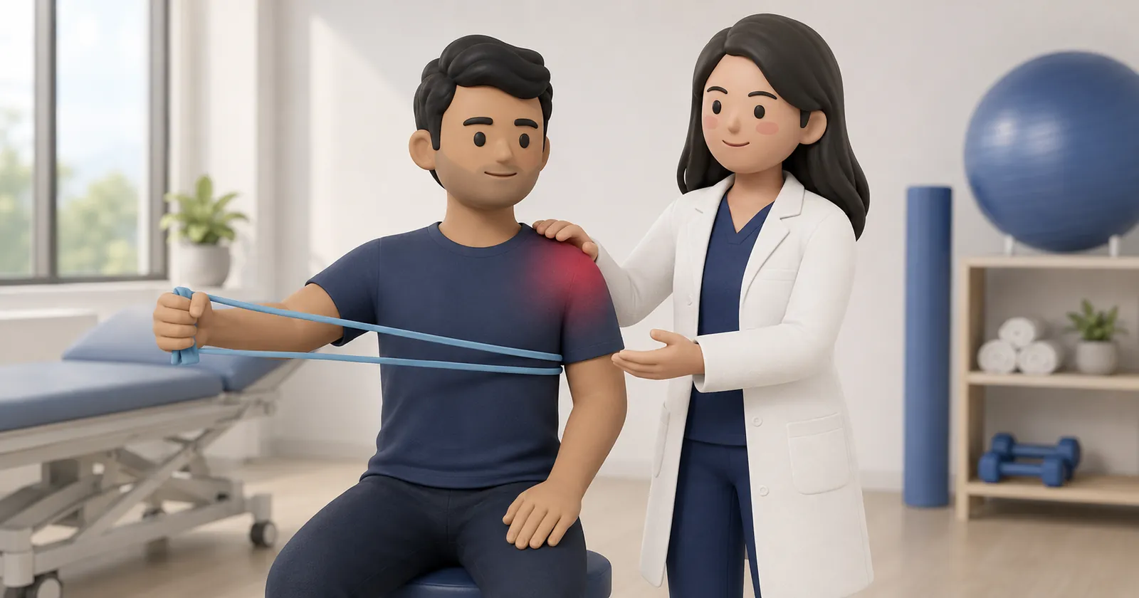
- Phase 3: Progressive Strengthening (Weeks 13 to 20): Light resistance band internal and external rotations with elbow at side, progressing to prone Y-and-T scapular strengthening and light dumbbell presses.
- Phase 4: Advanced Plyometrics and Sports (Months 6+): Rebounder ball catches, overhead sports drills, and heavy manual labor clearance once strength symmetry exceeds 85 percent.
Bathing and Sleeping Realities in Indian Homes
Sleeping flat supine frequently causes severe post-operative night pain due to gravitational posterior sag of the humerus.
Physiotherapists instruct patients to sleep in a 45-degree recliner chair or prop their back up with pillows in bed, supporting the operated elbow with a folded towel.
Bathing must be performed using a sponge wash while wearing a waterproof sling until sutures are removed.
Frequently Asked Questions
What is an abduction pillow sling and why is it necessary?
An abduction sling holds the arm 15 to 30 degrees away from the side of the body. This takes tension off the repaired supraspinatus tendon footprint and preserves capillary microcirculation to the healing repair.
How long does it take for the tendon to firmly re-attach to the bone?
Histological studies show that early collagen bridging begins by 6 to 8 weeks, but true structural enthesis maturation and tensile load capacity require 6 to 12 months.
Why is the shoulder so stiff after the sling comes off?
Temporary stiffness is an intentional consequence of protecting the repair. Once the sling is removed at week 6, physiotherapy safely and progressively restores full motion without endangering the tendon.
Related condition guides


Related recovery guides & articles
BookPhysio.in Editorial Team
The BookPhysio.in editorial team writes and maintains this content, checking it against reliable clinical sources. Content follows our medical review policy: general information only, and no page claims a clinical review until a named registered physiotherapist has signed it off.
Read our medical review policy