
Reverse Total Shoulder Arthroplasty: Biomechanics, Deltoid Rehab and Precautions
In short
Reverse Total Shoulder Arthroplasty inverts joint anatomy for massive rotator cuff tears. Learn deltoid recruitment, dislocation precautions, and recovery milestones.
In this guide (4 sections)
The Inverted Biomechanics of Reverse Total Shoulder Arthroplasty
Traditional anatomic shoulder replacement requires an intact, functional rotator cuff to depress and stabilize the humeral head against the glenoid socket. In cuff tear arthropathy (massive, irreparable rotator cuff tears with secondary glenohumeral arthritis), an anatomic prosthesis fails rapidly because the unconstrained humeral head migrates superiorly.
Reverse Total Shoulder Arthroplasty (rTSA), developed by Paul Grammont in 1985, completely inverts the normal anatomy: a spherical glenosphere is fixed to the glenoid bone, and a concave polyethylene socket is mounted onto the proximal humerus.
Grammont Biomechanical Principles and Deltoid Dominance
The reverse design fundamentally changes the muscle physics of the shoulder girdle.
| Biomechanical Parameter | Anatomic Shoulder Joint | Reverse Shoulder Joint (rTSA) | Functional Consequence | |
|---|---|---|---|---|
| Center of Rotation (COR) | Located within the anatomical humeral head | Shifted medially (onto glenoid surface) and distally | Lengthens the deltoid muscle lever arm by 20 to 30 percent. | |
| Primary Motor Driver | Supraspinatus initiates; deltoid assists elevation | Anterior and middle deltoid drives full elevation alone | Compensates 100% for absent rotator cuff muscles. | |
| Joint Stability Mechanism | Dynamic rotator cuff concavity-compression | Semi-constrained semi-spherical mechanical cup | Intrinsic stability against superior humeral migration. | |
| External Rotation Capacity | Driven by infraspinatus and teres minor | Severely limited unless teres minor or latissimus is intact | Rehab focuses on functional reaching; external rotation remains modest. |
Critical Dislocation Precautions and Phased Rehabilitation
Unlike an anatomic shoulder which dislocates anteriorly in abduction and external rotation, the reverse prosthesis is vulnerable to dislocation in a completely different movement vector.
- The Vulnerable Dislocation Position: Dislocation occurs in combined shoulder adduction, extension, and internal rotation (reaching behind the back to tuck in a shirt or wipe after toileting). This combination pushes the humeral cup out of the glenosphere.
- Phase 1: Protection (Weeks 0 to 6): Sling worn 24/7 except for elbow/wrist pumps. Passive forward flexion in the scapular plane capped at 90 to 100 degrees. Passive external rotation limited to 20 to 30 degrees. No active shoulder movement.
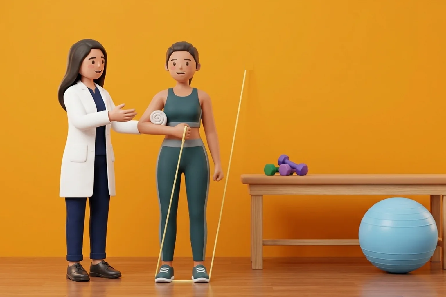
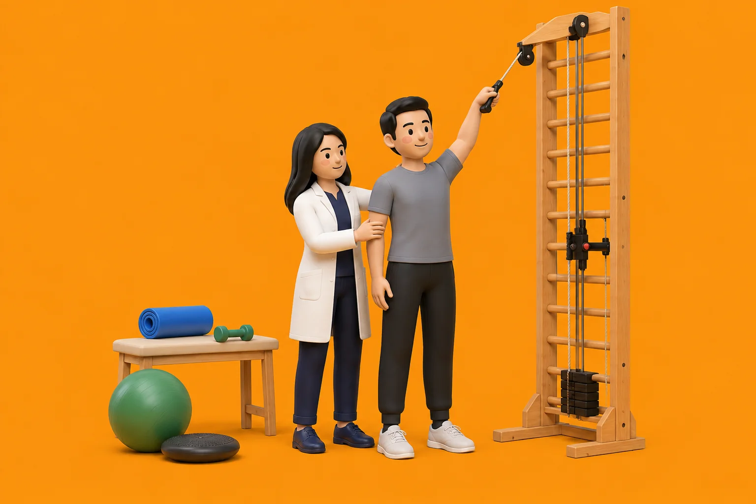
- Phase 2: Deltoid Re-Education (Weeks 6 to 12): Discontinue sling. Begin supine active-assisted forward elevation. Progress to seated active elevation, training the anterior deltoid using short lever arms (bent elbow) to minimize joint shear.
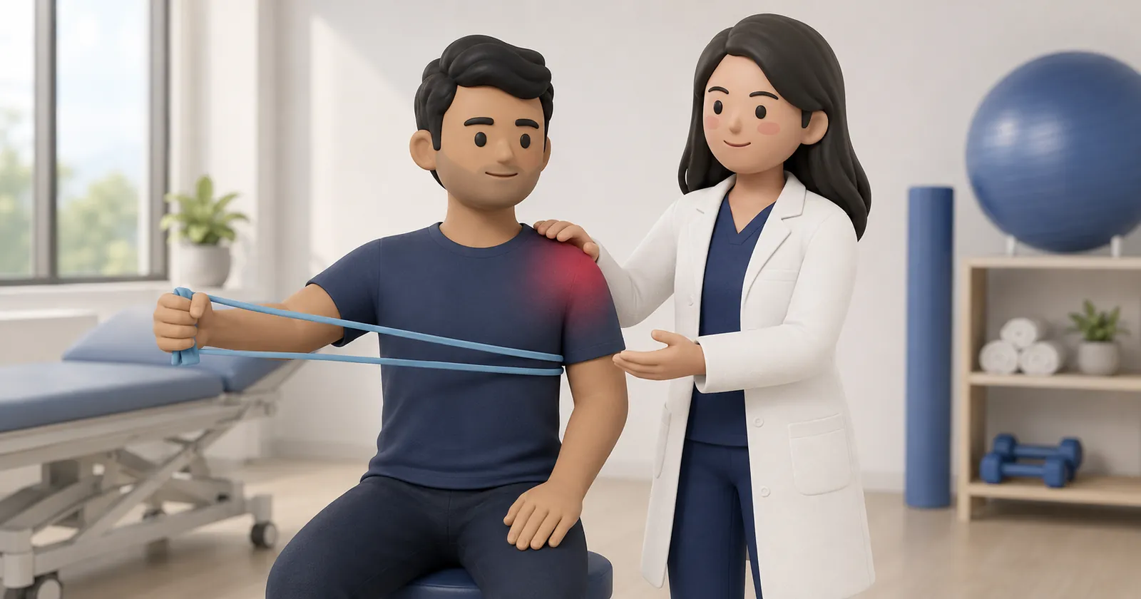
- Phase 3: Strengthening and Independence (Week 12+): Progressive light resistance bands for deltoid and periscapular stabilizers. Lifelong lifting limit: 5 to 7 kg for repetitive tasks.
Frequently Asked Questions
Can a patient regain full overhead reach after reverse shoulder surgery?
Most patients achieve active forward elevation between 120 and 140 degrees, which is sufficient for high kitchen reaching, washing hair, and dressing. True 180-degree reach is rare.
What is scapular notching in rTSA?
Scapular notching is mechanical impingement between the medial border of the humeral cup and the inferior neck of the scapula during arm adduction. Modern lateralized glenosphere designs have significantly reduced this complication.
How do patients manage personal hygiene without reaching behind their back?
Because reaching behind the back to the gluteal cleft is restricted to prevent dislocation, patients use long-handled toileting aids, handheld bidet sprays, or adapt side-reaching techniques.
Related condition guides
Related recovery guides & articles
BookPhysio.in Editorial Team
The BookPhysio.in editorial team writes and maintains this content, checking it against reliable clinical sources. Content follows our medical review policy: general information only, and no page claims a clinical review until a named registered physiotherapist has signed it off.
Read our medical review policy