
Suboccipital Release: Trigeminocervical Complex Desensitization and Cervicogenic Pain
In short
Cervical afferents converge directly onto trigeminal sensory neurons in the brainstem; suboccipital myofascial release unloads the trigeminocervical complex to abort headache.
In this guide (4 sections)
The Anatomy and Neurobiology of the Trigeminocervical Complex (TCC)
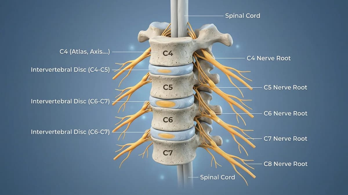
Cervicogenic headache and chronic suboccipital pain represent classic models of referred neurogenic distress. The anatomical foundation of this phenomenon is the Trigeminocervical Complex (TCC), located in the upper cervical spinal cord (C1 to C3 levels) and continuous with the caudal trigeminal nucleus (pars caudalis) in the medulla.
Primary nociceptive afferents from the upper three cervical spinal nerves (innervating the atlanto-occipital joint, atlanto-axial joint, C2-C3 zygapophyseal joints, and suboccipital musculature) converge directly upon the same second-order projection neurons that receive sensory inputs from the ophthalmic division (V1) of the trigeminal nerve. When suboccipital muscles (rectus capitis posterior major and minor, obliquus capitis superior and inferior) develop hypertonic spasm, sustained nociceptive bombardment sensitizes the TCC, causing the brain to misinterpret upper neck irritation as frontal, retro-orbital, or temporal head pain.
Upper Cervical Musculature and Clinical Diagnostic Differentiation
| Suboccipital Muscle Group | Anatomical Attachments & Innervation | Referred Pain Pattern on Palpation | Targeted Manual & Movement Therapy | |
|---|---|---|---|---|
| Rectus Capitis Posterior Major | Spinous process of Axis (C2) to inferior nuchal line of occiput; suboccipital nerve | Deep aching pain radiating over parietal skull to forehead | Fingertip ischemic compression at nuchal line; craniocervical flexion | |
| Rectus Capitis Posterior Minor | Posterior tubercle of Atlas (C1) to occiput; attaches to myodural bridge | Direct traction on spinal dura mater; intense throbbing retro-orbital pain | Sustained suboccipital traction to release tension on the myodural bridge | |
| Obliquus Capitis Inferior | Spinous process of C2 to transverse process of C1; suboccipital nerve | Periorbital ache, intense temporal pain; limits C1-C2 cervical rotation | C1-C2 Flexion-Rotation Test (FRT); manual sustained natural apophyseal glides (SNAGs) | |
| Obliquus Capitis Superior | Transverse process of C1 to occiput; suboccipital nerve | Band-like tension encircling temporal and occipital cranium | Lateral suboccipital soft-tissue glide and occipito-atlantal long-axis distraction |
Standardized Clinical Rehabilitation Protocols and Manual Release
- Sustained Manual Suboccipital Cranio-Cervical Decompression: Position the patient supine; cup your curved fingertips directly under the inferior nuchal line, allowing the weight of the patient's cranium to sink gently into your finger pads; apply gentle cephalad traction for 3 to 5 minutes to release deep fascial tension and relax the myodural bridge.
- C1-C2 Flexion-Rotation Mobilization (SNAG Protocol): Fully flex the patient's cervical spine to lock out the lower cervical segments (C3-C7), then gently rotate the head to the left and right; a difference of greater than 10 degrees between sides confirms C1-C2 hypomobility, treated with localized Mulligan apophyseal glides.
- Deep Cervical Flexor (Longus Colli/Capitis) Activation Drills: Prescribe supine craniocervical flexion training (gentle nodding action) using a pressure biofeedback unit inflated to 20 mmHg; guide the patient to progressively hold pressures from 22 to 30 mmHg for 10 seconds without recruiting superficial sternocleidomastoid muscles.
Frequently Asked Questions
Why does tightness at the very base of my skull cause severe throbbing behind my eyes?
The nerves from the top three joints of your neck meet and cross paths with your facial nerves in a relay station inside your brainstem (the trigeminocervical complex). When your deep neck muscles cramp up, your brain confuses the signals and thinks the pain is coming from behind your eyes or across your forehead.
What is the 'myodural bridge', and why does neck tension cause brain headaches?
Tiny connective tissue fibers directly link the small muscles at the base of your skull (rectus capitis posterior minor) to the protective lining of your spinal cord and brain (the dura mater). When these neck muscles are chronically tight, they physically tug on this lining, triggering headaches.
How often should I do the gentle chin-tuck exercises at my desk?
Perform 5 to 10 slow, gentle chin nods every hour while sitting at your computer. Imagine holding a ripe peach between your chin and throat without squishing it. This strengthens your deep neck stabilizers and prevents your skull muscles from tightening up.
Related condition guides

Related recovery guides & articles
BookPhysio.in Editorial Team
The BookPhysio.in editorial team writes and maintains this content, checking it against reliable clinical sources. Content follows our medical review policy: general information only, and no page claims a clinical review until a named registered physiotherapist has signed it off.
Read our medical review policy