
Autonomic Dysreflexia in T6 and Above: Identifying Hypertensive Crises
In short
Pathophysiology, early clinical recognition, triggers, and dangerous hypertensive complications of autonomic dysreflexia in individuals with spinal cord injuries at or above T6.
In this guide (3 sections)
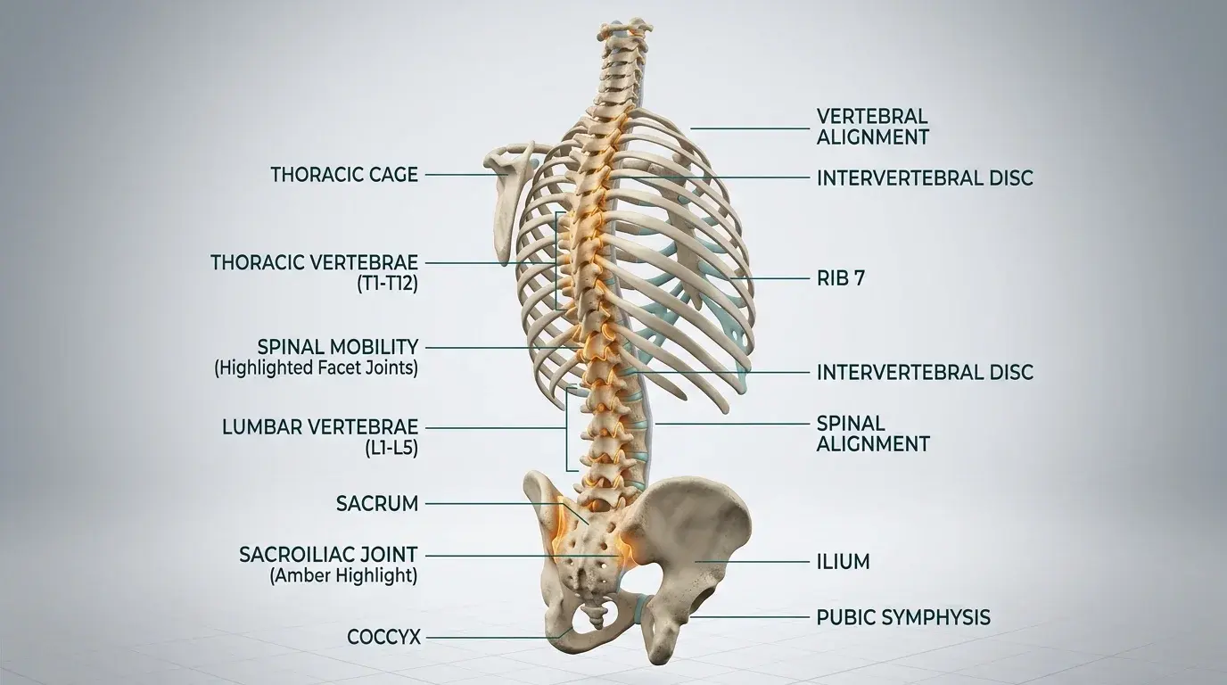
Autonomic dysreflexia (AD) is a potentially life-threatening medical emergency occurring in individuals with spinal cord injury at or above the sixth thoracic vertebra (T6). It is characterized by an uninhibited, massive sympathetic nervous system reflex discharge triggered by noxious or non-noxious stimuli below the level of the neurological lesion. If unrecognized and untreated, the resulting severe paroxysmal hypertension can precipitate intracranial hemorrhage, retinal detachment, seizures, cardiac arrhythmias, and death.
Pathophysiology of Sympathetic Hyperactivation in T6 and Above Lesions
Sensory afferent signals, primarily conveyed by spinothalamic tracts and dorsal columns from visceral organs or skin below the lesion, enter the spinal cord and trigger profound reflex excitation of preganglionic sympathetic neurons located in the intermediolateral cell column (T1 to L2). This sympathetic surge releases massive amounts of noradrenaline, causing intense peripheral and splanchnic arteriolar vasoconstriction. In neurologically intact individuals, baroreceptors in the carotid sinus and aortic arch detect the acute hypertension and stimulate medullary centers to send descending inhibitory signals to dampen sympathetic outflow. However, in cord lesions at T6 and above, descending inhibitory signals cannot pass the damaged cord segments. The brainstem can only attempt compensatory bradycardia via the intact vagus nerve (cranial nerve X), creating a dramatic mismatch of cranial parasympathetic vasodilation and caudal sympathetic vasoconstriction.
| Body Region | Autonomic Response | Observed Clinical Signs | Patient-Reported Symptoms | |
|---|---|---|---|---|
| Above Level of Injury (Parasympathetic) | Uninhibited vagal and cranial parasympathetic activity attempt to counteract hypertension. | Profuse facial flushing, forehead diaphoresis, nasal congestion, pupil constriction (miosis). | Pounding bitemporal or occipital headache, blurred vision, anxiety, cognitive slowing. | |
| Below Level of Injury (Sympathetic) | Unchecked massive sympathetic noradrenergic arteriolar vasoconstriction. | Pale, cold, clammy skin, piloerection (goosebumps), systemic arteriolar constriction. | Chills, shivering sensations, abdominal cramping, tight chest discomfort. | |
| Systemic Hemodynamic Profile | Severe paroxysmal hypertension with reflex vagal response. | Systolic BP increases >20 to 40 mmHg above baseline; paradoxical bradycardia (heart rate <60 bpm). | Sensation of impending doom, cardiac palpitations, dyspnea. |
Etiological Triggers and Systematic Bedside Triage
Over 85 percent of autonomic dysreflexia episodes originate from genitourinary triggers, predominantly acute bladder distension caused by a kinked, blocked, or overfilled catheter, or acute urinary tract infection (UTI). The second most common cause (10 to 12 percent) is gastrointestinal, involving fecal impaction, rectal vault distension, or hemorrhoidal thrombosis. Other triggers include ingrown toenails, restrictive clothing, tight shoe straps, pressure ulcers, and acute abdominal pathology.
Rapid Autonomic Dysreflexia Bedside Diagnostic Triage Protocol
Objective: Systematically identify and document the onset of autonomic dysreflexia in an acute rehabilitation or home environment within 60 seconds.
- 1Immediately obtain an arterial blood pressure reading using a calibrated manual sphygmomanometer or automated monitor.
- 2Compare current systolic blood pressure to the patient known baseline (tetraplegic baseline is typically low, around 90-100 mmHg).
- 3Observe the face, neck, and chest for cutaneous flushing, profuse sweating, and conjunctival injection.
- 4Inspect the limbs below the lesion for pallor, cool extremities, and prominent piloerection.
- 5Palpate the suprapubic area immediately to assess for acute bladder distension or a blocked urinary drainage bag.
Domestic Realities and Caregiver Education in Indian Homes
In Indian home care environments, tetraplegic patients are frequently cared for by family members or domestic attendants who have never received formal training on autonomic dysreflexia. A common catastrophic mistake occurs when family members see a patient with a pounding headache and profuse sweating, assume the patient is having a stroke or fainting spell, and lay them flat in bed. Laying a patient supine during autonomic dysreflexia increases intracerebral blood pressure and drastically elevates stroke risk. Every Indian caregiver must be taught that head-up sitting is mandatory.
Life-Threatening Hypertensive Sequelae and Surveillance
Failure to resolve autonomic dysreflexia rapidly can result in catastrophic vascular rupture. Blood pressures exceeding 200/120 mmHg in a patient whose baseline is 90/60 mmHg represent a severe medical crisis.
- Intracerebral Hemorrhage: Acute pressure waves rupture fragile intracranial arterioles, causing focal hemorrhagic stroke or subarachnoid bleed.
- Cardiac Arrhythmias: Excessive vagal tone combined with severe ventricular afterload induces heart blocks, severe bradycardia, or cardiac arrest.
- Pulmonary Edema: Massive splanchnic vasoconstriction shifts blood volumes rapidly into the central circulation, causing acute cardiogenic pulmonary edema.
Patients with cervical and high thoracic spinal cord injuries normally maintain a baseline blood pressure of 90/60 to 100/70 mmHg. A blood pressure reading of 130/85 mmHg in these patients represents significant hypertension and may signal active autonomic dysreflexia.
Frequently Asked Questions
Why does autonomic dysreflexia only occur in injuries at or above T6?
The major sympathetic splanchnic vascular outflow originates between thoracic levels T6 and L2, innervating the vast vascular bed of the abdominal viscera. Injuries above T6 leave this massive vascular bed isolated from descending brainstem inhibitory pathways, allowing unconstrained vasoconstriction.
Can a person with a complete T4 injury develop autonomic dysreflexia during an uneventful bowel routine?
Yes. Digital rectal stimulation, insertion of suppositories, or hard stool stretching the rectal ampulla commonly triggers an autonomic dysreflexia crisis. Using topical 2 percent lignocaine jelly 5 minutes prior to digital rectal stimulation reduces this sensory reflex trigger.
Do patients in India need a doctor referral to begin specialized neuro-physiotherapy?
Under the National Commission for Allied and Healthcare Professions (NCAHP) Act 2021, registered physiotherapists are recognized as primary contact practitioners. You can consult a licensed neuro-physiotherapist directly without a physician prescription. At BookPhysio.in, every practitioner is verified, and you pay directly per session without hidden fees or platform markups.
Related condition guides



Related recovery guides & articles
BookPhysio.in Editorial Team
The BookPhysio.in editorial team writes and maintains this content, checking it against reliable clinical sources. Content follows our medical review policy: general information only, and no page claims a clinical review until a named registered physiotherapist has signed it off.
Read our medical review policy