
Complex Pelvic Pain Syndrome Following Healed Pelvic Ring Injuries
In short
Managing chronic complex pelvic pain after healed pelvic ring trauma. Master sacroiliac micromotion, pelvic floor hypertonicity, and neuromodulation.

In this guide (5 sections)
While orthopaedic trauma surgeons celebrate radiological union following complex pelvic ring disruptions, up to 30 to 50 percent of patients experience persistent, disabling chronic symptoms recognized as Post-Traumatic Complex Pelvic Pain Syndrome (CPPS). Persisting long after bone fractures consolidate, this complex syndrome arises from a combination of persistent sacroiliac joint micromotion, asymmetric pelvic ring load transmission, hypertonic pelvic floor muscle dysfunction, and central sensitization of the lumbosacral plexus. Effective physical rehabilitation requires an advanced approach combining joint stabilization, pelvic floor myofascial down-training, and sensory desensitization.
Domestic and Cultural Indian Realities
In Indian society, chronic pelvic pain following traumatic injuries carries severe psychological and social stigma, particularly for young adult survivors. Persistent pain during prolonged sitting, deep groin aching, and discomfort during intimate relations or defecation are frequently suffered in silence due to cultural embarrassment. In multi-generational households, family members may perceive complaints of pain after healed X-rays as psychological weakness. Physiotherapists provide compassionate, objective validation, explaining the neuro-muscular basis of post-traumatic pain and prescribing private home exercises.
Biomechanical Malunion, Pudendal Neuralgia, and Pelvic Floor Spasm
Even minor residual pelvic displacement (such as a 5 mm vertical step-off or 1 cm posterior rotational asymmetry) fundamentally alters lumbopelvic load transfer, concentrating abnormal shearing moments across the sacroiliac joint and pubic symphysis. Simultaneously, traumatic traction on the pudendal nerve (S2-S4) or sacral plexus during initial injury leads to neuropathic allodynia and hyperalgesia. In response to chronic pain, the pelvic floor musculature (levator ani, coccygeus, obturator internus) enters a state of chronic protective hypertonicity, creating internal trigger points that refer pain to the perineum, groin, and lower back.
| Clinical Domain | Pain Etiology | Pathophysiological Mechanism | Clinical Presentation | Physiotherapy Rehabilitation Protocol |
|---|---|---|---|---|
| Sacroiliac Micromotion | Ligamentous laxity and residual rotational malunion | Sharp buttock pain radiating to posterior thigh; pain on rolling | Pelvic stabilization belt; isometric transverse abdominis and gluteal bracing | |
| Pelvic Floor Hypertonicity | Protective muscle spasm of levator ani and obturator internus | Deep perineal aching; pain worsened by sitting; dyspareunia | Diaphragmatic breathing; reverse Kegel relaxation drills; pelvic floor down-training | |
| Pudendal Nerve Sensitization | Post-traumatic traction injury and perineural fibrosis | Burning, lancinating pain in perineum and genitalia; sitting intolerance | Pudendal nerve desensitization; contoured cutout seating cushions; TENS | |
| Central Sensitization | Widespread dorsal horn excitability and pain amplification | Generalized tenderness; kinesiophobia; sleep disruption | Pain neuroscience education; graded motor imagery; aerobic walking pacing |
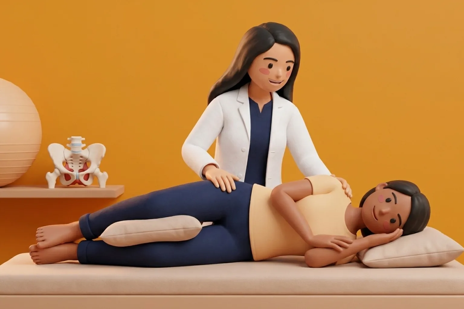
Pelvic Floor Down-Training and Diaphragmatic Breath Integration
Traditional pelvic floor exercises (Kegels) that focus on contracting the muscles are strictly contraindicated in hypertonic pelvic pain, as they exacerbate muscle spasm. Instead, rehabilitation prioritizes down-training and relaxation: the patient is taught diaphragmatic breathing combined with the reverse Kegel technique, consciously visualizing the pelvic floor muscles dropping and opening like an umbrella during inhalation.
Supported Supine Diaphragmatic Reverse Kegel Relaxation
Objective: Release chronic hypertonic spasm in the levator ani and obturator internus muscles through respiratory coupling.
- 1Position the patient supine on a firm bed with knees bent, feet flat, and calves resting on a soft bolster.
- 2Place one hand over the upper chest and the other over the lower abdomen just above the pubic bone.
- 3Instruct the patient to breathe in slowly through the nose, allowing the abdomen to expand gently outward.
- 4At peak inhalation, cue the patient to visualize the muscles between the sitting bones softening and dropping.
- 5Exhale gently through relaxed lips without forcing, maintaining the sensation of pelvic floor openness.
If the patient experiences new or rapidly progressive urinary retention, fecal incontinence, saddle anesthesia (numbness over the buttocks and groin), or severe foot weakness, evaluate immediately for cauda equina syndrome or late screw migration.
Long-Term Surveillance and Complication Prevention
Surveillance tracks pain intensity using the Visual Analogue Scale (VAS) and evaluates quality of life using the Pelvic Floor Distress Inventory (PFDI-20). Long-term pacing strategies prevent flare-ups.
- Pelvic Floor Distress Inventory Audits: Administer the PFDI-20 questionnaire monthly to objectively evaluate improvements in urinary, colorectal, and pelvic organ prolapse distress domains.
- Seated Tolerance Mapping: Document continuous sitting tolerance in minutes on contoured memory-foam versus gel cutout cushions to track functional workstation readiness.
- Graded Aerobic Conditioning Logging: Monitor daily step counts using a pedometer, advancing walking targets by 10 percent weekly while avoiding boom-and-bust pain flare cycles.
Frequently Asked Questions
Why do I still have severe pain in my pelvic area even though the doctor says my bones have healed?
Bone healing on X-rays is only one part of recovery. The severe accident also stretched your pelvic floor muscles, nerves, and ligaments. Your pelvic muscles can get stuck in a tight, protective spasm that pinches nerves and causes deep, aching pain even after the bones are solid.
Should I do Kegel exercises to strengthen my pelvic floor after a pelvic fracture?
No. Doing standard tightening Kegel exercises will make hypertonic pelvic pain worse. Your pelvic muscles are already clenched too tightly in spasm. Physiotherapy teaches you reverse Kegels and deep breathing to relax and soften these muscles, relieving pressure on irritated nerves.
Do I need a doctor referral to begin home physiotherapy after complex trauma or external fixation?
Under the National Commission for Allied and Healthcare Professions (NCAHP) Act 2021, patients in India enjoy direct access to registered physiotherapists without requiring a mandatory doctor referral. At BookPhysio.in, you connect with verified clinical physiotherapists and pay directly per session without advance packages, hidden platform fees, or long-term contracts.
Related condition guides

Related recovery guides & articles
BookPhysio.in Editorial Team
The BookPhysio.in editorial team writes and maintains this content, checking it against reliable clinical sources. Content follows our medical review policy: general information only, and no page claims a clinical review until a named registered physiotherapist has signed it off.
Read our medical review policy