
Master Lumbar Spinal Stenosis and Neurogenic Claudication Guide
In short
Explore the definitive clinical master guide to lumbar spinal stenosis, neurogenic claudication physics, differential diagnosis, and rehabilitation in India.
In this guide (4 sections)
Lumbar spinal stenosis (LSS) and its primary symptomatic manifestation, neurogenic claudication, represent one of the most prevalent and debilitating musculoskeletal disorders affecting aging adults across the globe. Characterized by a progressive narrowing of the central thecal canal, lateral recesses, and intervertebral neural foramina, spinal stenosis transforms routine ground ambulation into an exhausting ordeal. As life expectancy rises in India, millions of senior citizens find their physical independence, community mobility, and emotional vitality compromised by posture-dependent leg cramping, heaviness, and balance instability. This master clinical guide synthesizes the complete pathophysiology, diagnostic differential testing, domestic biomechanics, and evidence-based physical rehabilitation protocols for managing lumbar spinal stenosis, providing a comprehensive, compassionate blueprint for lasting functional recovery.
Bazaar Walking Fatigue, Mandi Vegetable Bags, and Bus Stop Standing Limitations
In Indian society, the functional impact of spinal stenosis permeates every dimension of daily existence. Older adults typically struggle with everyday cultural rituals: walking through bustling neighborhood sabzi mandis, standing stationary at bus stops, waiting in temple darshan lines, or preparing daily meals at kitchen counters. Carrying heavy grocery bags and standing upright force the spine into extension, closing the spinal canal and triggering severe bilateral calf cramping within minutes. Seniors frequently discover that leaning over fruit vendor carts, resting on park benches, or stooping forward provides immediate relief. Understanding that these postures reflect dynamic canal physics rather than physical weakness empowers families to implement supportive adaptations, restoring mobility across the neighborhood.
Dynamic Canal Physics, Microvascular Ischemia, and Postural Kinematics
The central hallmark of lumbar spinal stenosis is its dynamic, posture-dependent nature. In healthy spines, the lumbar canal maintains ample reserve capacity. In degenerative stenosis, disc height collapse, facet joint arthrosis, and ligamentum flavum hypertrophy reduce midsagittal canal dimensions below ten millimetres. When standing or walking upright, physiological lumbar lordosis narrows the central canal by up to sixteen percent, compressing the thecal sac and elevating epidural venous pressure. Because venous congestion prevents arterial capillary inflow, cauda equina nerve roots suffer transient ischemic conduction blocks, producing pseudoradicular leg heaviness and paresthesia. Transitioning into forward lumbar flexion widens the central canal by up to twenty-four percent, immediately restoring microvascular perfusion and clearing neurogenic symptoms.
Multimodal Rehabilitation: Decompression, Pacing, and Kinetic Stabilization
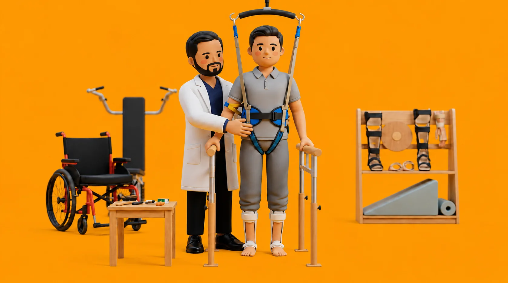
Modern physical rehabilitation for spinal stenosis follows a multimodal, evidence-based paradigm. Therapy rejects obsolete passive machine treatments in favor of active, patient-centered conditioning. First, flexion-biased stabilization exercises train the deep abdominal wall and gluteal muscles to eliminate compensatory lumbar hyperextension. Second, walking interval pacing calculates individual claudication thresholds, allowing patients to expand total daily step counts through structured, sub-symptom walking bouts. Third, stationary recumbent cycling and aquatic therapy provide rigorous cardiovascular conditioning in an unloaded, decompressed posture. Fourth, assistive devices: such as dual trekking poles and rollators, transfer upper-body weight away from the spine, allowing seniors to participate confidently in community life.
Master Triple Lumbar Decompression and Core Integration Sequence
Objective: Combine supine flexion decompression, posterior pelvic tilting, and core activation into a daily master routine
- 1Lie on your back on a firm bed or mat with both knees bent, feet flat, and a thin pillow supporting your neck.
- 2Perform a posterior pelvic tilt by gently tightening your lower abdomen and pressing your lower back firmly flat against the floor.
- 3While maintaining this flat contact, draw both knees up toward your chest, clasping your hands behind your thighs.
- 4Hold this deep flexion stretch for thirty seconds, taking slow diaphragmatic breaths to decompress the spinal canal.
- 5Slowly lower your feet back to the mat one at a time and perform 8 gentle alternating heel slides while maintaining core contact with the mat.
| Clinical Feature | Diagnostic Indicator & Pathophysiology | Evidence-Based Rehabilitation Strategy |
|---|---|---|
| Central Canal vs Vascular Claudication | Neurogenic: relief with flexion (sitting, stooping); normal pedal pulses; positive van Gelderen test | Flexion stabilization, stationary cycling, walking interval pacing; avoid straight-leg extension |
| Posture-Dependent Canal Physics | Extension narrows canal by 16 percent; flexion expands canal by up to 24 percent | Williams flexion protocols, supported hip hinging, patla footrest while standing, rollator gait |
| Secondary Kinetic Chain Deficits | Hip flexor contractures, gluteus medius weakness (Trendelenburg), multifidus fatty atrophy | Thomas stretch modifications, resisted band clamshells, mini-squats, and protein optimization |
| Autonomous Legal & Pricing Framework | Direct access authorized under NCAHP Act 2021; autonomous evaluation and red flag triage | Transparent pay-per-session pricing (₹600 to ₹1,200); avoid coercive upfront package sales |
If you experience sudden bowel or bladder retention, involuntary leakage, numbness between your thighs around the saddle area, or sudden inability to lift your feet when walking, seek emergency medical care immediately. These indicate acute cauda equina compression requiring urgent intervention.
Frequently Asked Questions
What is the most important thing I can do at home to manage my lumbar spinal stenosis?
The single most effective strategy is practicing walking interval pacing. Instead of walking until severe pain forces you to stop, take short walking breaks before pain starts, resting in a seated forward-leaning posture for one to two minutes. Combining this with daily flexion exercises and stationary cycling keeps your spinal canal open and protects your nerves.
Do I need a doctor referral to consult a spinal stenosis physiotherapist on BookPhysio.in?
BookPhysio.in does not require a doctor referral. Under the National Commission for Allied and Healthcare Professions (NCAHP) Act 2021, patients across India enjoy direct access to autonomous registered physiotherapists. Through BookPhysio.in, you connect directly with verified clinicians and pay per session with transparent fees, completely free from advance package commitments, commission markups, or platform subscription charges.
Related condition guides



Related recovery guides & articles
BookPhysio.in Editorial Team
The BookPhysio.in editorial team writes and maintains this content, checking it against reliable clinical sources. Content follows our medical review policy: general information only, and no page claims a clinical review until a named registered physiotherapist has signed it off.
Read our medical review policy