
Neuromuscular Electrical Stimulation (NMES) in Critically Ill Patients
In short
Prescribe NMES for ICU muscle preservation. Review stimulus parameters, electrode placement, membrane excitability, and clinical protocols in India.
In this guide (2 sections)
Neuromuscular Electrical Stimulation (NMES) has emerged as an indispensable physiotherapy intervention capable of inducing repetitive skeletal muscle contractions in deeply sedated, comatose, or uncooperative critically ill patients. By delivering transcutaneous electrical currents directly to terminal motor nerve branches, NMES bypasses absent central voluntary motor drive. This modality stimulates muscle protein synthesis, enhances microvascular capillary perfusion, and preserves cross-sectional muscle architecture during the acute phases of critical illness immobilization.
Cellular Bioenergetics and Muscle Membrane Excitability in the ICU
Critical illness accelerates myofibrillar breakdown via the hyperactive ubiquitin-proteasome system and downregulates the Akt/mTOR protein synthesis pathway. Prolonged immobility also induces muscle membrane inexcitability through resting membrane depolarization and sodium channel inactivation. Applying standardized biphasic symmetrical electrical pulses produces rhythmic mechanical tension within myofibers, stimulating local secretion of insulin-like growth factor-1 (IGF-1), downregulating atrogin-1 and MuRF-1, and attenuating muscle atrophy even when the patient cannot voluntarily participate in physical exercise.
| Target Muscle Group | Pulse Frequency & Width | Duty Cycle (On / Off) | Electrode Dimensions & Placement | |
|---|---|---|---|---|
| Quadriceps Femoris | 35 to 50 Hz, 350 to 400 microsec | 5 seconds on / 15 seconds off (1:3) | Large rectangular pads (5x10 cm) over motor points of vastus lateralis and rectus femoris | |
| Gastrocnemius-Soleus | 30 to 45 Hz, 300 to 350 microsec | 6 seconds on / 18 seconds off (1:3) | Dual square electrodes over proximal medial and lateral muscle bellies | |
| Peroneal / Tibialis Anterior | 35 to 40 Hz, 250 to 300 microsec | 4 seconds on / 12 seconds off (1:3) | Proximal electrode below fibular neck, distal electrode over lower third anterior tibia | |
| Biceps & Triceps Brachii | 30 to 40 Hz, 250 to 300 microsec | 5 seconds on / 15 seconds off (1:3) | Dual circular or rectangular electrodes centered directly over mid-muscle bulk |
Standardized Electrode Application and Intensity Titration
Before applying electrodes, the therapist inspects skin integrity, cleans the cutaneous surface with saline wipes to remove sebum, and clips excessive hair. Peripheral anasarca and subcutaneous fluid accumulation commonly increase electrical impedance in critically ill patients, requiring larger conductive gel pads. Intensity is gradually titrated upward until a visible, palpable muscle contraction is elicited without provoking sympathetic tachycardia, hypertension, or painful grimacing.
Bilateral Quadriceps Motor Point NMES with Telemetry Monitoring
Objective: Preserve patellofemoral mobility and maintain knee extensor bulk in deeply sedated patients.
- 1Ensure the patient is supine with knees supported in 15 degrees of flexion using a firm foam roll.
- 2Place the proximal electrode over the upper third of the rectus femoris and the distal electrode over the vastus medialis motor point.
- 3Connect lead wires securely and set the stimulator to a symmetrical biphasic waveform at 45 Hz.
- 4Slowly increase current amplitude in small 2-milliamp increments while observing the muscle belly for rhythmic twitching.
- 5Achieve a visible tetanic muscle contraction and maintain the automated stimulation cycle for thirty minutes.
Domestic Recovery in India and Multigenerational Support
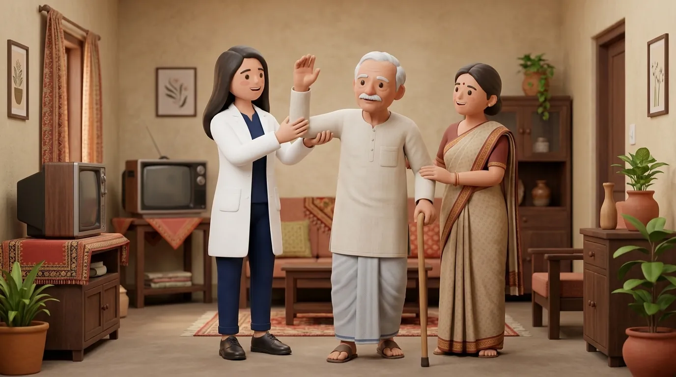
Following discharge to an Indian domestic environment, portable battery-operated NMES devices can bridge the transition to active standing. In multigenerational families, adult children or family attendants can be trained by visiting physiotherapists to apply electrodes safely, adjust intensity dials, and store lead wires properly, ensuring consistent daily rehabilitation while the patient regains voluntary motor recruitment.
Long-Term Trajectory and Muscle Architecture Recovery
Integrating NMES during acute ICU immobilization significantly preserves muscle cross-sectional area and accelerates the transition to active exercise. Longitudinal follow-up demonstrates that patients who receive protocolized NMES achieve independent standing and unassisted bed-to-chair transfers significantly earlier than non-stimulated controls, establishing a strong foundation for full functional independence.
Contraindications include demand cardiac pacemakers, implanted cardioverter-defibrillators, active lower extremity deep vein thrombosis, open skin wounds, and severe local cutaneous infections. Never place electrodes across the carotid sinuses or anterior neck.
Can NMES cause muscle damage or elevated serum creatine kinase in ICU patients?
When calibrated to submaximal clinical frequencies (30 to 50 Hz) with appropriate duty cycles (1:3 ratio), NMES does not cause myofiber necrosis or pathological elevations in serum creatine kinase. The low-frequency stimulation preserves muscle architecture without inducing metabolic overload.
Do I need a doctor referral to consult a home physiotherapist in India?
Under the National Commission for Allied and Healthcare Professions (NCAHP) Act 2021, patients in India enjoy direct access to registered physiotherapists without requiring a mandatory doctor referral. At BookPhysio.in, you consult verified physiotherapists and pay directly per session without hidden fees or platform commissions.
Related condition guides



Related recovery guides & articles
BookPhysio.in Editorial Team
The BookPhysio.in editorial team writes and maintains this content, checking it against reliable clinical sources. Content follows our medical review policy: general information only, and no page claims a clinical review until a named registered physiotherapist has signed it off.
Read our medical review policy