
Wound Breakdown and Sural Nerve Neuroma Post-Surgical Achilles Repair
In short
Learn to recognize and manage surgical wound breakdown, skin flap necrosis, and sural nerve entrapment complications following Achilles repair.
In this guide (4 sections)
While open surgical repair provides robust mechanical apposition of ruptured Achilles tendon ends, it carries a well-documented risk of soft-tissue envelope complications. The posterior heel and distal lower leg possess a uniquely fragile cutaneous anatomy, characterized by a thin subcutaneous layer, minimal muscular coverage, and an angiosome watershed blood supply dependent on terminal branches of the peroneal and posterior tibial arteries. Surgical wound breakdown, ranging from superficial skin edge maceration and wound dehiscence to full-thickness flap necrosis and deep peritendinous infection, occurs in two to ten percent of open cases. Simultaneously, the proximity of the sural nerve along the lateral margin of the incision renders it vulnerable to surgical traction, accidental puncture, or entrapment within scar tissue, resulting in painful stump neuromas and chronic lateral foot dysesthesias.
Indian Climatic Humidity, Dust Infiltration, and Dressing Moisture Hazards
In India, postsurgical wound care demands heightened vigilance due to environmental factors. High atmospheric humidity and intense seasonal heat induce profuse sweating beneath cast padding and orthotic liners, creating warm, moist conditions ideal for bacterial proliferation. In urban and rural residences alike, airborne dust and environmental particulates can penetrate loose dressings. Furthermore, accidental water splashing during bucket bathing or wet floor cleaning can saturate the cotton dressing. Patients must inspect surgical incisions daily under bright lighting, ensure all dressings remain bone-dry, and clean the skin surrounding the incision with sterile alcohol-free antiseptic wipes. Never apply traditional herbal pastes, turmeric powders, or unverified oils to surgical incisions.
Microvascular Thrombosis, Full-Thickness Necrosis, and Sural Neuroma Biology
The vascular biology of the posterior lower leg explains its vulnerability to wound complications. The midline longitudinal incision transects transversely oriented dermal capillary networks, relying entirely on longitudinal microvascular loops. Excessive retraction during surgery, hematoma formation beneath the skin flaps, or tight closure under tension compromises local perfusion pressure, inducing capillary thrombosis and full-thickness dermal necrosis. If bacteria penetrate the necrotic skin barrier, infection can track along suture lines into the tendon core, threatening tendon viability. Meanwhile, trauma to the sural nerve disrupts the perineurium. Regenerating axonal sprouts fail to find distal Schwann cell basal lamina tubes, forming a disorganized, tangled mass of unmyelinated axons and fibrous tissue: a traumatic terminal neuroma that displays extreme mechanosensitivity to light touch.
Early Triage Protocols: Managing Dehiscence and Sural Nerve Desensitization
Clinical management of wound complications depends on the depth of tissue involvement. Superficial wound separation without tendon exposure is managed conservatively with aggressive non-weight-bearing, oral culture-directed antibiotics, and specialized negative pressure wound therapy (NPWT) or silver-impregnated hydrofiber dressings. If deep tendon exposure occurs, urgent plastic surgical consultation for local rotational flaps (such as a reverse sural neurofasciocutaneous flap) or free tissue transfer is mandatory. For sural nerve irritability, physiotherapists initiate graded neurodynamic mobilization, lateral foot desensitization techniques using progressive textures (silk, cotton, terrycloth), and transcutaneous electrical nerve stimulation (TENS) to suppress neuropathic wind-up.
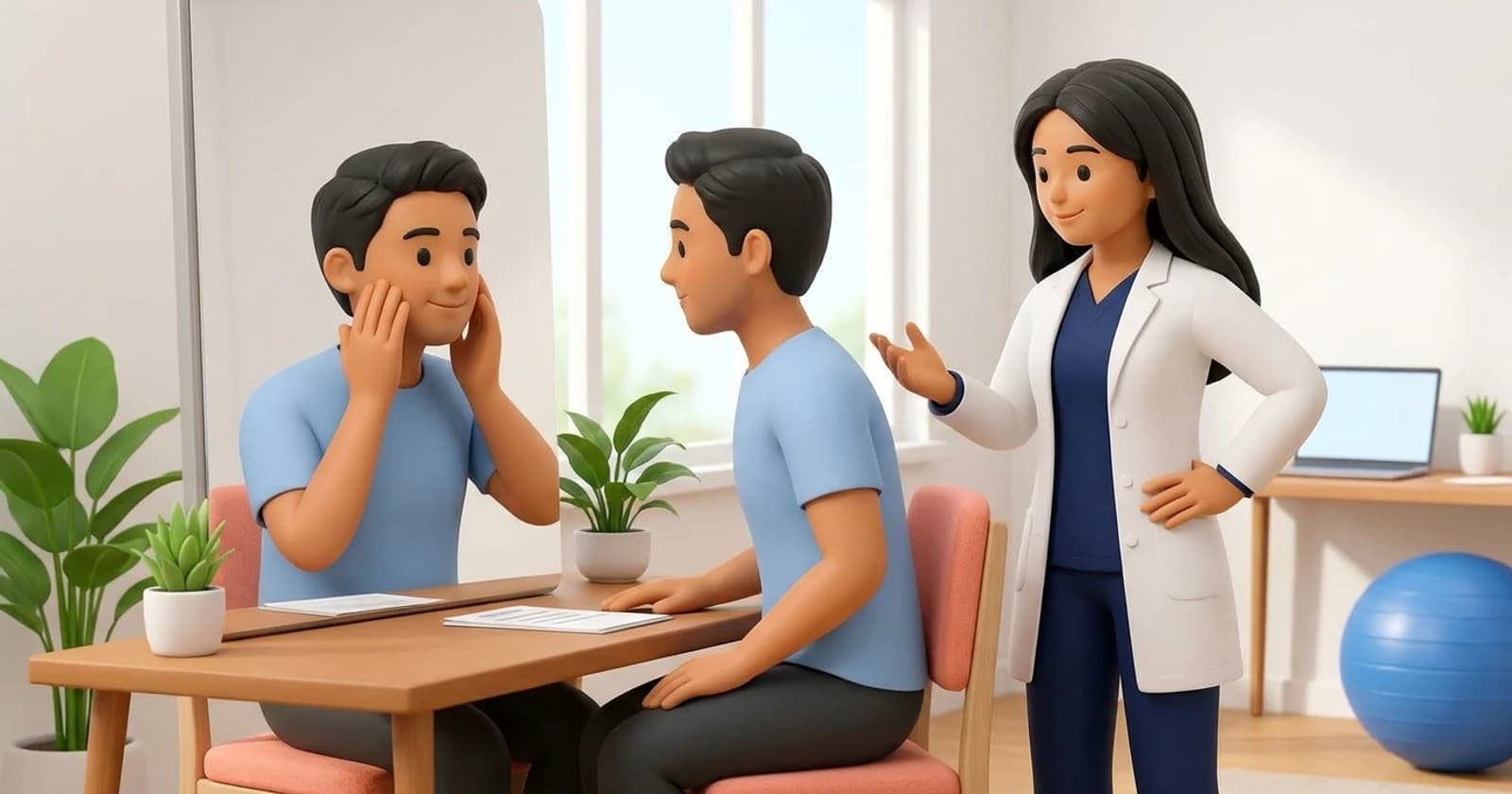
Seated Sural Nerve Gliding and Lateral Foot Desensitization
Objective: Encourage longitudinal excursion of the sural nerve through its soft-tissue interface while reducing neuropathic hypersensitivity
- 1Sit upright on a flat treatment plinth or bed with your legs extended straight in front of you.
- 2Keep your injured ankle in a relaxed, comfortable position, avoiding sharp dorsiflexion.
- 3Gently point your toes downward (plantarflexion) while turning your foot slightly inward (inversion) to lightly tension the lateral nerve pathway.
- 4Simultaneously extend your neck to look upward toward the ceiling, reducing tension at the spinal cord origin.
- 5Now slowly lower your chin toward your chest while allowing your foot to relax back to neutral, repeating this gentle gliding oscillation.
| Complication Category | Clinical Presentation Signs | Underlying Pathophysiology | Required Medical Action |
|---|---|---|---|
| Superficial Skin Necrosis | Dark eschar or separation along incision margins; tendon fully covered | Dermal edge microvascular ischemia without deep fascial breakdown | Debridement of non-viable margins; non-adherent silver dressings; close monitoring |
| Deep Wound Dehiscence | Gapping incision exposing underlying glistening tendon or suture material | Full-thickness dermal necrosis with secondary deep bacterial inoculation | Immediate surgical debridement; culture-guided antibiotics; possible tissue flap |
| Sural Nerve Neuroma | Sharp burning pain along lateral heel; hyperpathia; positive Tinel sign | Traction injury or scar entrapment of sural nerve fibers with axonal sprouting | Neurodynamic mobilization, neuropathic medications, local anesthetic nerve blocks |
If you observe yellow pus, expanding redness, foul odor, or visible white suture or tendon fibers through a separated incision, contact your operating orthopaedic surgeon immediately. Tendon drying and deep infection can cause complete structural liquefaction of the repair. Seek emergency evaluation without delay.
Frequently Asked Questions
Why does the outside edge of my foot feel numb or tingly after Achilles surgery?
The sural nerve courses directly along the lateral side of the Achilles tendon, providing skin sensation to the outside edge of your foot and little toe. During surgery, this delicate nerve is gently retracted or can become swollen from local surgical inflammation, causing temporary numbness or tingling (neuropraxia). In most patients, normal sensation gradually returns over three to six months as nerve swelling subsides and local tissues soften.
Do I need a doctor referral for Achilles rupture rehabilitation on BookPhysio.in?
BookPhysio.in does not require a doctor referral. Under the National Commission for Allied and Healthcare Professions (NCAHP) Act 2021, patients across India enjoy direct access to autonomous registered physiotherapists. Through BookPhysio.in, you connect directly with verified clinicians and pay per session with transparent fees, completely free from advance package commitments, commission markups, or platform subscription charges.
Related condition guides



Related recovery guides & articles
BookPhysio.in Editorial Team
The BookPhysio.in editorial team writes and maintains this content, checking it against reliable clinical sources. Content follows our medical review policy: general information only, and no page claims a clinical review until a named registered physiotherapist has signed it off.
Read our medical review policy