
ARDS Post-Discharge Rehabilitation: Prone Recovery, Diffusion Capacity and Exertional Hypoxia
In short
Surviving Acute Respiratory Distress Syndrome (ARDS) leaves persistent lung fibrosis and diffusion impairments. Learn post-discharge pacing, oxygen titration, and progressive exercise.
In this guide (5 sections)
Pathophysiology and Long-Term Sequelae of ARDS
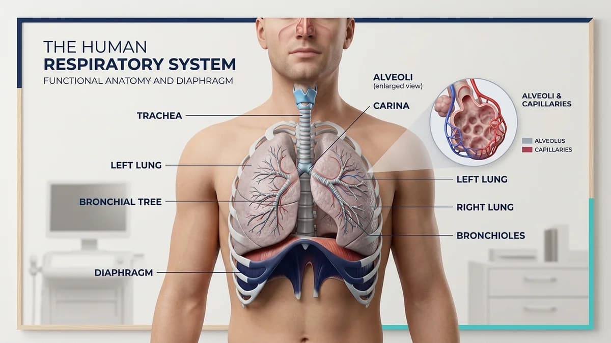
Acute Respiratory Distress Syndrome (ARDS) is characterized by diffuse alveolar-capillary membrane disruption, severe proteinaceous pulmonary edema, surfactant inactivation, and extensive microvascular thrombosis, typically triggered by viral pneumonia, sepsis, or aspiration.
While acute intensive care emphasizes lung-protective mechanical ventilation (low tidal volume of 4 to 6 mL/kg) and prone positioning, surviving patients frequently endure persistent alveolar fibrotic remodeling.
Pulmonary function testing 6 to 12 months post-discharge frequently shows marked reductions in Diffusing Capacity of the Lungs for Carbon Monoxide (DLCO), while spirometry (FVC and FEV1) may normalize, creating a clinical picture of severe exertional hypoxia despite near-normal resting spirometry.
Clinical Assessment and Exertional Desaturation Patterns
ARDS survivors display rapid desaturation during low-to-moderate metabolic activity due to ventilation-perfusion mismatch and diffusion limitation across thickened alveolar septa.
| Assessment Parameter | Normal Healthy Baseline | Post-ARDS Expected Value | Clinical Rehabilitation Consequence | |
|---|---|---|---|---|
| 1. DLCO (% Predicted) | > 80 percent | 40 to 60 percent (impaired diffusion) | Oxygen saturation drops rapidly with exercise; supplemental oxygen required. | |
| 2. 6-Minute Walk Test (6MWT) | 500 to 700 meters | 250 to 380 meters | Severe functional exercise limitation; high cardiopulmonary work. | |
| 3. Exertional SpO2 Drop | Stable SpO2 >= 96 percent | Desaturation to < 88 percent within 2-3 mins | Mandatory pulse oximetry pacing; stop walking when SpO2 < 88%. | |
| 4. Diaphragm Thickness Fraction | > 30 percent thickening on ultrasound | < 15 percent (diaphragm atrophy) | Accessory neck muscle overuse; paradoxical abdominal breathing. |
Structured Pulmonary Rehabilitation Protocol
Exercise training must be calibrated to avoid oxygen desaturation below 88 percent, utilizing interval training paradigms rather than continuous sustained aerobic loads.
Pacing allows capillary transit time in the pulmonary circulation to equilibrate, preventing severe systemic hypoxia and pulmonary hypertension flares.
- Interval Treadmill or Corridor Walking: Prescribe 2 to 3 minute bouts of walking at a Borg dyspnea rating of 3 to 4 (moderate), interspersed with 1 to 2 minutes of seated rest, accumulating 20 minutes total.
- Continuous Pulse Oximetry Titration: Titrate portable oxygen concentrators to maintain resting and ambulatory SpO2 strictly at or above 90 percent throughout all exercise sessions.
- Upper Extremity Resistance Training: Light resistance band exercises (seated bicep curls, chest press) to strengthen shoulder girdle musculature without causing accessory respiratory fatigue.
- Thoracic Expansion and Posture Realignment: Gentle spine extension and pectoralis major stretching to counteract restrictive chest wall splinting resulting from prolonged prone ICU positioning.
Indian Urban Air Pollution Considerations
ARDS survivors living in heavily polluted Indian metros (Delhi, Mumbai, Kolkata) face acute exacerbation risks from ambient PM2.5 and PM10 particles that irritate damaged alveolar membranes.
Physiotherapists instruct patients to perform structured exercise indoors in rooms equipped with HEPA air filtration during high smog months (October to February).
Frequently Asked Questions
How long does lung tissue take to heal after ARDS?
Fibroproliferative resolution continues for 12 to 18 months. Many patients experience substantial gains in DLCO and functional walking capacity through consistent pulmonary rehabilitation during this window.
Is it dangerous if oxygen saturation drops to 85 percent during exercise?
Yes. Exertional desaturation below 88 percent strains the right ventricle and can precipitate cardiac arrhythmias or pulmonary vasoconstriction. Exercise must be paused immediately until SpO2 recovers to >= 90 percent.
Why did prone positioning help in the ICU?
Prone positioning improves ventilation-perfusion matching by relieving dorsal lung compression from the heart and abdominal contents, opening collapsed alveoli.
Related condition guides

Related recovery guides & articles
BookPhysio.in Editorial Team
The BookPhysio.in editorial team writes and maintains this content, checking it against reliable clinical sources. Content follows our medical review policy: general information only, and no page claims a clinical review until a named registered physiotherapist has signed it off.
Read our medical review policy