
Symphysis Pubis Dysfunction (SPD): Pubic Symphysis Gap, Waddling Gait & Functional Transfers
In short
Severe shooting pain at the front pubic bone makes walking feel like your pelvis is splitting. Discover evidence-based physiotherapy solutions for symphysis pubis dysfunction.

In this guide (5 sections)
Anatomical Pathophysiology of the Pubic Symphysis
The symphysis pubis is an amphiarthrodial fibrocartilaginous joint connecting the left and right superior pubic rami, reinforced by superior, inferior (arcuate), anterior, and posterior pubic ligaments. Under non-pregnant conditions, physiologic joint separation is 4 to 5 millimeters with minimal translation.
During pregnancy, hormonal loosening combined with fetal gravitational weight widens this gap to 8 to 9 millimeters. If widening exceeds 10 to 12 millimeters (diastasis of the pubic symphysis) or if asymmetric vertical shearing occurs during single-leg stance, sharp localized pain radiates into the groin, inner thighs, and lower abdomen.
Biomechanics of the 'Waddling' Gait
Patients with SPD instinctively adopt a wide-based, lateral-swaying 'waddling' gait to minimize the single-leg support phase and avoid shearing the inflamed pubic fibrocartilage.
While this gait is a natural protective compensation, excessive lateral sway places tremendous strain on the gluteus medius and tensor fasciae latae, leading to secondary trochanteric bursitis and myofascial fatigue.
| Clinical Test | Procedure | Positive Indication | Reliability | |
|---|---|---|---|---|
| Pubic Bone Palpation | Direct gentle pressure applied over superior pubic disc | Sharp, shooting local pain remaining > 5 seconds | High specificity for pubic inflammation | |
| Patrick's / FABER Test | Hip flexion, abduction, and external rotation in supine | Reproduces anterior groin/pubic pain rather than posterior SIJ | Standard diagnostic orthopedic battery | |
| Stork / Gillette Test | Single-leg stance with contralateral hip flexion | Failure to stabilize; acute click or shear at pubic bone | Assesses dynamic pelvic ring stability |
Functional Ergonomics and Pain-Free Daily Transfers
The overarching rule in SPD rehabilitation is: 'Keep your knees together.' Any movement that widens the thighs creates direct vertical and rotational torque across the pubic joint.
Simple functional adjustments significantly reduce daily mechanical aggravation.
- Car Entry and Exit: Sit down sideways onto the car seat first, then swivel both legs and knees inward together as a single unit.
- Stair Climbing Modifications: Take stairs one step at a time, leading with the less painful leg going up and the symptomatic leg going down.
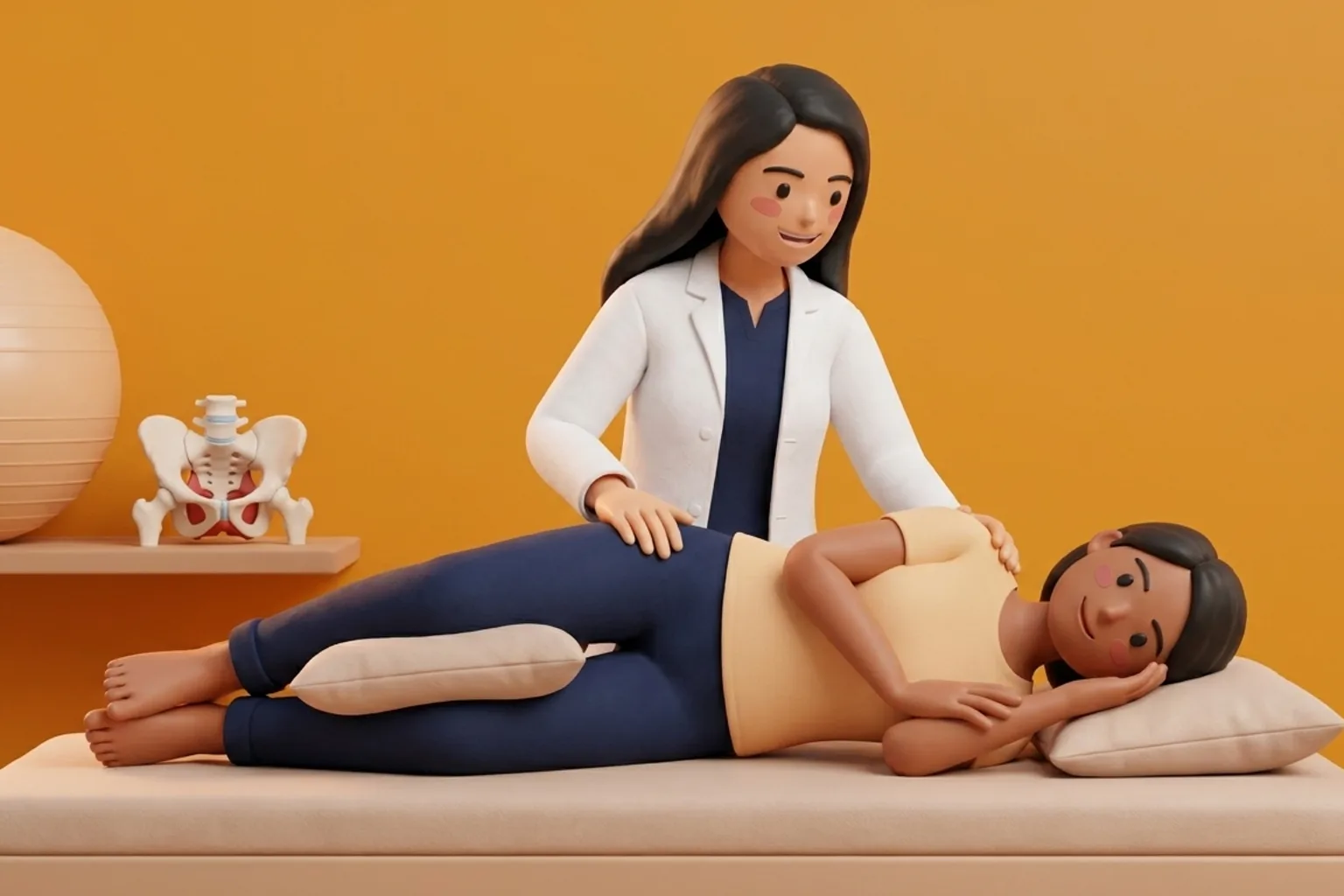
- Getting Out of Bed: Roll onto your side with knees glued together, push up with your arms while swinging both lower legs over the edge simultaneously.
Labor and Delivery Positioning Strategies
Women with severe SPD should avoid standard lithotomy positions (lying flat with legs forced wide open into stirrups), which place maximum stretch on the pubic symphysis. Ideal birth positions include all-fours (quadruped), side-lying with the upper leg supported on a peanut ball, or kneeling forward over a birthing ball.
Frequently Asked Questions
Can I have a normal vaginal delivery if I have severe SPD?
Yes. Most women with SPD have safe, uncomplicated vaginal deliveries. Your physiotherapist can measure your comfortable pain-free abduction range and note it in your birth plan so your healthcare team avoids over-stretching your legs during labor.
Why does my pubic bone click or pop when I turn in bed?
The click is caused by microscopic joint translation and air cavitation within the widened, mobile symphysis pubis joint as your weight shifts from one pelvic half to the other.
What pain relief modalities are safe for SPD during pregnancy?
Ice packs applied over the pubic bone for 15 minutes, antenatal trochanteric support belts, targeted isometric adductor squeeze exercises, and gentle water therapy are safe and effective.
Related condition guides

Related recovery guides & articles
BookPhysio.in Editorial Team
The BookPhysio.in editorial team writes and maintains this content, checking it against reliable clinical sources. Content follows our medical review policy: general information only, and no page claims a clinical review until a named registered physiotherapist has signed it off.
Read our medical review policy