
Stroke Rehabilitation at Home: Transfer Techniques, Gait Retraining, and Arm Recovery Guide
In short
Home-based stroke rehabilitation harnesses the brain neuroplastic potential during the critical post-acute recovery window. Learn safe caregiver transfer techniques, spasticity management, and functional gait retraining.
In this guide (5 sections)
A stroke (cerebrovascular accident) is a life-altering medical emergency that strikes over 1.8 million individuals across India each year. When blood supply to a region of the brain is interrupted by an ischemic clot or hemorrhagic vessel rupture, neural tissue suffers rapid oxygen deprivation, resulting in contralateral hemiplegia (weakness or paralysis on the opposite side of the body), sensory deficits, spatial neglect, and impaired motor coordination. Following initial acute medical stabilization in a hospital ICU, the most crucial phase of recovery begins: intensive, home-based neurorehabilitation.
The biological foundation of stroke recovery is neuroplasticity: the remarkable capacity of the central nervous system to reorganize its neural architecture, form new synaptic connections, and recruit undamaged cortical pathways to take over functions previously managed by injured brain regions. The golden biological window for maximal neuroplastic adaptation occurs within the first 3 to 6 months post-stroke. During this critical window, repetitive, high-frequency, task-oriented physiotherapy delivered in the familiar domestic setting of an Indian home drives vastly superior functional recovery compared to bedbound isolation.
1. Managing Hemiplegic Muscle Tone: Flaccidity vs Spasticity
Following a stroke, muscular tone typically evolves through predictable neurological stages, historically outlined by Signe Brunnstrom. In the initial acute stage, the affected limb often presents with Flaccidity (complete hypotonia), where muscles feel limp, heavy, and unresponsive. During this flaccid stage, protecting the paretic joints is life-critical: the paralyzed shoulder is exceptionally vulnerable to inferior subluxation under the pull of gravity.
As cortical shock subsides, Spasticity (velocity-dependent hypertonia and hyperreflexia) emerges. In the upper extremity, spasticity characteristically locks the limb into an abnormal Flexor Synergy Pattern: internal shoulder rotation, adduction against the rib cage, elbow flexion, forearm pronation, and clenched fisting of the wrist and fingers. In the lower extremity, an Extensor Synergy Pattern dominates: hip extension and adduction, rigid knee extension, and plantarflexion with inversion of the ankle (equinovarus foot drop).
Effective physiotherapy does not simply fight spasticity with passive stretching; it utilizes prolonged inhibitory positioning, slow rotational trunk mobilization, and functional task-oriented movement to channel primitive reflexes into controlled, volitional motor actions.
2. Safe Caregiver Transfer Techniques: Protecting the Shoulder
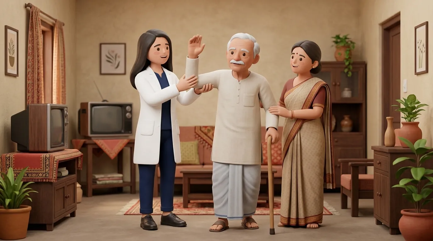
In Indian households, family members and domestic attendants are the bedrock of daily care. However, well-meaning caregivers frequently cause severe iatrogenic trauma by pulling the stroke patient up from a bed or chair by their paralyzed arm. This traction force stretches the flaccid rotator cuff ligaments, resulting in painful shoulder subluxation and triggering agonizing Complex Regional Pain Syndrome (Shoulder-Hand Syndrome).
| Transfer Stage | Dangerous Caregiver Error | Correct Safe Biomechanical Technique | Clinical Rationale |
|---|---|---|---|
| Bed-to-Chair Pivot Transfer | Pulling the patient upward by their paretic arm or grabbing under the armpit | Position chair at 45° to strong side; support patient around waist or use a transfer belt | Completely eliminates downward traction on the vulnerable glenohumeral joint capsule |
| Rolling in Bed (Log Rolling) | Dragging the patient across the bedsheet from the hemiplegic side | Clasp patient hands together (strong hand holding weak hand); bend knees; roll toward weak side as a unit | Inhibits flexor spasticity and stimulates sensory awareness of the neglected hemiplegic side |
| Sit-to-Stand Transition | Allowing patient to pull themselves up using the caregiver neck or clothing | Place both patient feet flat on floor; shift weight forward ("nose over toes"); push from armrests | Promotes symmetrical bilateral quadriceps activation and normal vestibular postural reflexes |
| Arm Support in Wheelchair / Chair | Allowing the paretic arm to dangle off the side of the chair or wheelchair | Support the paretic forearm on a contoured lap tray or pillow in 30° abduction and neutral wrist | Prevents gravitational traction and keeps the humeral head safely centered in the glenoid fossa |
3. Three-Phase Home Stroke Rehabilitation Protocol
Structured home rehabilitation follows a progressive sequence: mastering bed mobility, progressing to dynamic sitting balance, and advancing to overground gait training.
Three-Phase Task-Oriented Home Stroke Recovery Protocol
Objective: Promote neuroplastic motor recovery, inhibit abnormal synergies, and rebuild independent functional mobility.
- 1Phase 1 (Bed Mobility and Pelvic Bridging): Lie supine with knees bent and feet flat on the bed. Clasp your hands together with interlaced fingers, pointing arms straight toward the ceiling. Squeeze your gluteal muscles and lift your pelvis off the bed into a bridge, holding for 5 seconds. Pelvic bridging develops the core and hip extensor control necessary for future walking.
- 2Phase 2 (Symmetrical Sit-to-Stand and Weight-Shifting): Sit on a firm dining chair with feet shoulder-width apart. Shift your body weight consciously toward the paretic leg until you feel equal weight distribution. Lean your chest forward over your knees, drive through both heels, and stand tall. Repeat for 3 sets of 8 repetitions under close supervision.
- 3Phase 3 (Task-Oriented Arm Reach and Constraint-Induced Drills): Sit at a dining table. Place both forearms on the table. Slide a folded hand towel across the table forward and backward using both arms together, opening and closing the paretic hand. Practice grasping light plastic cups and moving them across targets to stimulate corticospinal motor map reorganization.
- 4Overground Walking Progression: Begin walking with an appropriate assistive device (such as a broad-based quadripod cane or hemi-walker held in the non-paretic hand). Focus on heel-strike and symmetrical step length rather than walking speed.
4. Orthotics: Ankle-Foot Orthosis (AFO) and Slings
Foot drop (inability to dorsiflex the ankle during the swing phase of walking) is present in over 70% of stroke survivors, causing dangerous toe-dragging and compensatory circumduction (hiking the hip outward to clear the foot). Fitting a custom thermoplastic Ankle-Foot Orthosis (AFO) holds the ankle in 90 degrees of neutral dorsiflexion, preventing trips and falls on uneven household carpets or thresholds.
For the upper limb, an orthopaedic humeral cuff sling (such as a GivMohr sling) should be worn during standing and walking to support the weight of the flaccid arm, while allowing the arm to move freely during seated therapy.
Frequently Asked Questions
Can a stroke survivor recover motor function after the 6-month window in India?
Yes. While spontaneous biological recovery is most rapid in the first 3 to 6 months, neuroplasticity never completely closes. Clinical trials prove that stroke survivors can continue regaining muscle strength, fine motor coordination, and functional walking ability years after a stroke, provided they engage in intensive, repetitive, task-specific physiotherapy and progressive strength training rather than passive bed rest.
Why should caregivers never pull a stroke patient by their weak arm?
During the flaccid or hypotonic stage of a stroke, the rotator cuff muscles that normally hold the ball of the arm bone inside the shoulder socket are paralyzed. Pulling on the arm causes the humeral head to slip downward out of the socket (inferior subluxation), tearing the delicate joint capsule and triggering chronic, agonizing shoulder pain that severely impedes overall recovery.
What is the role of electrical stimulation in stroke hand recovery?
Neuromuscular Electrical Stimulation (NMES) and Functional Electrical Stimulation (FES) are valuable adjuncts in stroke rehabilitation. Delivering controlled electrical impulses to the radial nerve (wrist extensors) and deep peroneal nerve (ankle dorsiflexors) stimulates motor neuron firing, helps overcome sensory neglect, reduces extensor spasticity through reciprocal inhibition, and accelerates volitional movement.
How do you prevent bed sores and chest infections in bedridden stroke patients?
Execute a strict 2-hour positional log-rolling schedule: alternate the patient posture between back lying, right side-lying, and left side-lying to relieve pressure over the sacrum, greater trochanters, and heels. Deploy an alternating ripple pressure air mattress, and perform daily chest physiotherapy (assisted deep breathing and gentle back percussion) to mobilize lung secretions and prevent hypostatic pneumonia.
How does BookPhysio.in support home stroke recovery across Indian cities?
BookPhysio.in connects stroke survivors and families across India with verified neurological physiotherapists who specialize in home-based neurorehabilitation, caregiver transfer training, spasticity management, and gait re-education. Patients pay transparent per-session fees directly at each visit (₹600 to ₹2,000 for specialized home visits) with zero platform commission.
Related condition guides


BookPhysio.in Editorial Team
The BookPhysio.in editorial team writes and maintains this content, checking it against reliable clinical sources. Content follows our medical review policy: general information only, and no page claims a clinical review until a named registered physiotherapist has signed it off.
Read our medical review policy